Figures & data
Figure 1. Exocytosis of large secretory vesicles. A. Modality of exocytosis. Secretory vesicles may undergo exocytosis following three different modalities: (1) single fusion, (2) kiss and run, and (3) compound exocytosis. B. Diagram of a SGs acinus and organization of the APM. Acini are formed by pyramidal polarized epithelial cells that are in close contact and the APM (red) is shared by two cells and forms narrow canaliculi. The dashed boxes show two orthogonal enlargements of the apical area. The canaliculi have a diameter of 0.2‑0.3 µm and are separated from the basolateral membrane by tight junctions (green). The SCGs (blue) have a much larger diameter (1‑1.5 µm). When the fusion pore opens two compartments with different composition and membrane tension are interconnected (diagram on the right).
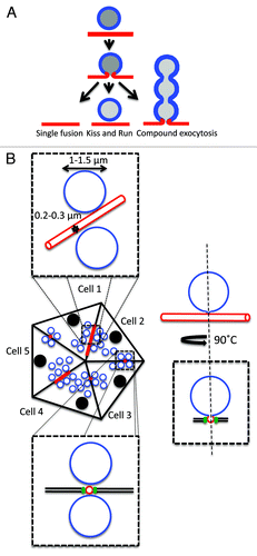
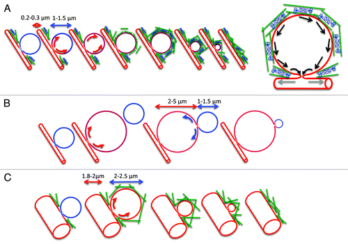
Figure 2. Role of the actomyosin complex in the gradual collapse of the secretory granules. A. SCGs (blue) fuse with the APM (red), the fusion pore opens, and membranes flow from the APM into the SCGs (red arrows). The difference in membrane tension may not favor the flow of the membranes toward the APM that would lead to the gradual collapse of the SCGs. The contractile activity of the actomyosin complex (actin, green rods; myosin II, blue) that assembles around the SCGs may push the membranes (black arrows) and/or dilate further the fusion pore (gray arrows) to facilitate the gradual collapse. B. In the absence of a functional actin cytoskeleton the SCGs expand forming large vacuoles (2‑5 µm). Membranes flow into the large vacuoles, which acquire the properties of the APM. Due to the lower membrane tension of the large vacuoles, the remaining SCGs gradually collapse without the need of a functional actomyosin complex. C. In the absence of a functional myosin II, the acinar canaliculi significantly expand in size (1.8‑2.0 µm). The SCGs fuse with the APM and slightly expand in size (2‑2.5 µm). Under these conditions the difference in membrane tension may be more favorable to a actomyosin-independent gradual collapse.