Figures & data
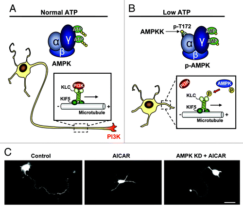
Figure 1. Schematic illustration depicting the mechanism of AMPK dependent polarity inhibition. (A) Under normal energy conditions AMPK exists in an unphosphorylated/inactive state and PI3K is transported to the neurite tip via a physical association with the kif5 cargo adaptor, KLC. The accumulation of PI3K at a single neurite tip promotes the signaling responsible for axon initiation and growth. (B) Under energy-lacking conditions, AMP binds to AMPK producing a conformational change in the kinase, allowing phospho-activation of AMPK by upstream kinases (AMPKK). AMPK-caused KLC phosphorylation dissociates PI3K, resulting in a loss of PI3K from the neurite tip and an inhibition of neuronal polarization. (C) Cultured hippocampal neurons are transfected with GFP for visualization. Control neuron shows typical single axon (left), which is missing in a neuron treated with AMPK activator AICAR (middle). Expression of kinase dead (KD) AMPK rescues polarity in the AICAR treated neuron. Scale bar = 20 μm.