Figures & data
Figure 1 Following mitosis, dorsal neural tube cells can reinsert into the neuroepithelium. (A) A neural epithelial cell labeled with γ-tubulin-GFP (green) and mCherry-tubulin (red) and indicated within the white box is shown in the time-lapse series below. (A′) As this cell (white dot) undergoes interkinetic nuclear migration and mitosis at the apical surface, the centrosome (white arrowhead) leaves the apical surface and travels basally (0:14). In the next time frame (0:21), the nucleus moves towards the apical domain and meets the duplicated centrosomes in a non-apical location (white and blue arrowheads), and then the centrosomes and the nucleus travel together to the apical surface, where cytokinesis occurs (completed by 0:56). Note that the plane of cell division separates the basal-most daughter cell (white dot) from the apical surface. Following cell division, the centrosome (blue arrowhead) of the apical-most daughter cell (blue dot) is found at the lumen, whereas the centrosome (white arrowhead) belonging to the basal-most daughter cell (white dot) is found at a non-apical position. As the time-series progresses, the cell tail with the non-apical centrosome (white arrowhead) returns to the apical surface (complete by 4:26). Scale bars are 10 µm. D = dorsal, nt = neural tube, nc = neural crest, ap = apical.
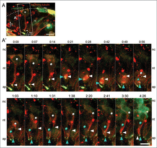
Figure 2 Neural crest emigration accompanied by filopodia and blebs. (A) The mem-EGFP-labeled neural epithelial cell indicated within the white box is shown in the time-lapse series below. (A′) Note that there are two different mem-EGFP-labeled cells close together within the field of view, a dorsal neural tube cell (red dot) and a neural crest cell (no dot). The neural epithelial cell (red dot) detaches from the lumen and migrates out of the neural tube. The formation of filopodia are indicated with red arrowheads and membrane blebs with red arrows. Scale bars are 10 µm.
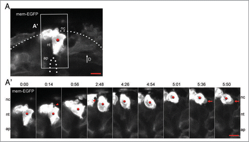
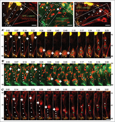
Figure 3 Examples of neural epithelial cells undergoing INM. (A–C) Neuroepithelial cells were labeled with GFP-α-catenin (green) and mCherry-tubulin (red). Cells within the white boxes are shown in the time-lapse series below. (A′) Prior to nuclear migration in an apical direction, the mCherry-tubulin signal in the cell tail is reduced (white arrowheads) and mCherry-tubulin enters the nucleus (white arrow, t = 0:21). The nucleus is then transported from a basal position to the apical surface, where the mitotic spindle forms (bright mCherry-tubulin signal) and cytokinesis occurs. The nuclei of the daughter cells (white and blue dots) are then transported basally. (B′) Before nuclear migration from the basal to apical surface, the mCherry-tubulin signal in the cell tail is reduced (white arrowheads) and mCherry-tubulin enters the nucleus (white arrow, t = 0:28). Mitotic spindle formation also occurs while the nucleus is in a basal position (bright mCherry-tubulin signal, t = 0:28). The nucleus is then transported to the apical surface where cytokinesis occurs. The daughter cell nuclei (white and blue dots) are subsequently shuttled to a basal position. (C′) Note the presence of two different mCherry-tubulin cell tails in the viewing field. Prior to nuclear migration from the basal to apical surface, the mCherry-tubulin signal in one cell tail is reduced (white arrowheads) and mCherry-tubulin enters the nucleus (white arrow, t = 0:28). Nuclear migration in the apical direction and spindle formation occurs at t = 0:35, but the nucleus does not make it all the way to the apical surface before cell division occurs (t = 0:56). After cytokinesis, the nuclei of the daughter cells (white and blue dots) are transported in a basal direction. Scale bars are 10 µm.