Figures & data
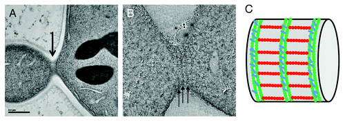
Figure 1. Electron microscopy characterization of bud neck septin filaments in S. cerevisiae. (A) Projection view of a 50 nm section from a dividing budding yeast. Grazing filaments are seen close to the membrane (arrow). (B) Single slice from a tomographic reconstruction of a grazing section of budding yeast. Circumferential filaments are indicated with arrows. (C) Schematic representation of septin filaments at the bud neck based on tomographic analysis, with circumferential filaments shown in green and axial filaments drawn in red.