Figures & data
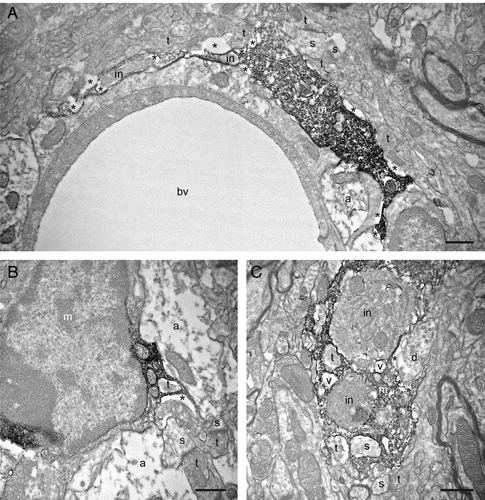
Figure 1. Putative microglial filopodial protrusions contacting synaptic elements in a mouse model of HIV-associated neurocognitive disorder (HAND). A-B: Filopodia from the same cell (shown by the arrows in A) are simultaneously targeting three asymmetrical synapses between axon terminals (t) and dendritic spines (s). C: A putative microglial process that contains a vacuole (v) is simultaneously touching a dendritic spine and a symmetrical synapse between an axon terminal and a dendrite (d). D: Another process juxtaposing a neuronal cell body (n) is contacting an axon terminal and an astrocytic process (a). m, putative microglia; ma, myelinated axon. Pockets of extracellular space are shown by the asterisks (*). Scale bars: 1μm.
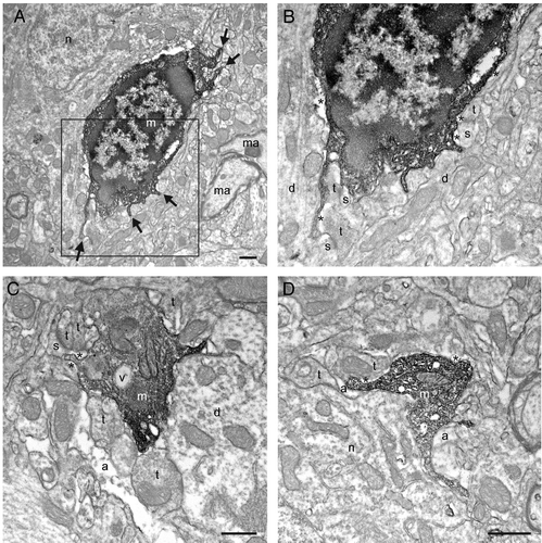
Figure 2. Additional examples of putative microglial filopodial protrusions contacting synaptic elements (A-D), proximal dendrites (B-C), a blood vessel (bv; C), and myelinated axons (D). Note that B and Care showing a magnified view of the putative microglia in A. Annotations as in . Scale bars: 1μm.
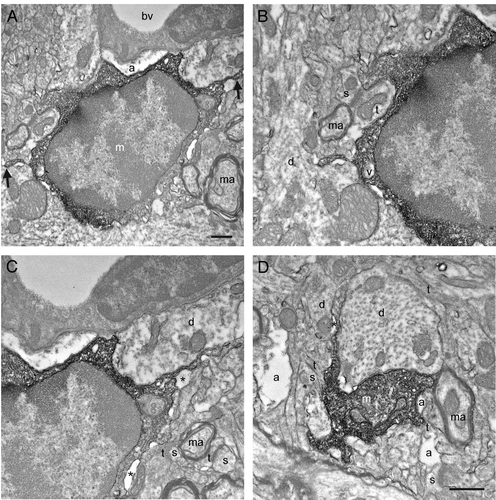
Figure 3. Examples of putative microglial cell body, large and small processes in the saline-injected controls (A-D) observed in direct juxtaposition with synaptic elements like dendritic spines (s) and axon terminals (t), as well as perisynaptic astrocytic processes (a). Note the absence of filopodial protrusions from the cell body in A, as during normal physiological conditions. Annotations as in and . Scale bars: 1μm.
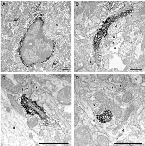
Figure 4. Filopodial protrusions displaying phagocytic inclusions (in) nearby their extremity (A) and closer to the cell body (B). In C, a putative proximal microglial process is containing several cellular inclusions, including two axon terminals (t), a dendritic spine (s) and two vacuoles (v). Note that the engulfed elements are typically surrounded by extracellular space, which suggests their ongoing proteolytic degradation. Other annotations as in , and . Scale bars: 1μm.