344
Views
3
CrossRef citations to date
0
Altmetric
Article Addendum

Dictyostelium Centrin B localization during cell cycle progression

, &
Pages 39-41 | Received 31 Jul 2009, Accepted 03 Aug 2009, Published online: 01 Jan 2010

Figures & data

Figure 1 Localization of DdCenB at three stages of the cell cycle. DdCenB is shown as gray shading, the darkness of which corresponds to the amount of DdCenB. N, nucleus; NE, nuclear envelope; C, centrosome; PM, plasma membrane.
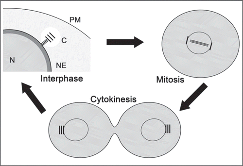
Figure 1 Localization of DdCenB at three stages of the cell cycle. DdCenB is shown as gray shading, the darkness of which corresponds to the amount of DdCenB. N, nucleus; NE, nuclear envelope; C, centrosome; PM, plasma membrane.

Figure 2 Cytokinesis defects as a result of DdCenB knock-out. (A) consists of images captured from time-lapse microscopy. The time of capture (minutes) for each frame is indicated in the lower right corners. The arrowheads point to the persistent cytoplasmic bridges observed in dividing cells. In (B) we see a wild-type cell dividing. Here the arrowhead points to a typical cleavage furrow. (C) is a dividing mutant cell stained with DA PI (DNA) and visualized by fluorescence/DIC microscopy. Arrows point to distorted nuclei in the furrow region. (D) shows a mutant cell, with a long cytoplasmic bridge, stained with DAPI. The arrows point to higher magnifications of the respective areas, and highlight the presence of DNA in the cytoplasmic bridge. D, fluorescence; D’, phase contrast. Bars in all cases correspond to 5 µm.
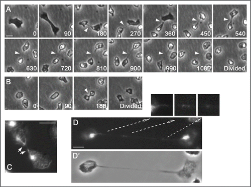
Figure 2 Cytokinesis defects as a result of DdCenB knock-out. (A) consists of images captured from time-lapse microscopy. The time of capture (minutes) for each frame is indicated in the lower right corners. The arrowheads point to the persistent cytoplasmic bridges observed in dividing cells. In (B) we see a wild-type cell dividing. Here the arrowhead points to a typical cleavage furrow. (C) is a dividing mutant cell stained with DA PI (DNA) and visualized by fluorescence/DIC microscopy. Arrows point to distorted nuclei in the furrow region. (D) shows a mutant cell, with a long cytoplasmic bridge, stained with DAPI. The arrows point to higher magnifications of the respective areas, and highlight the presence of DNA in the cytoplasmic bridge. D, fluorescence; D’, phase contrast. Bars in all cases correspond to 5 µm.