Figures & data
Figure 1 Domain Architecture of MICAL-family of proteins. FAD: Flavin-adenine dinucleotide binding domain, CH: Calponin Homology domain, LIM: LIM domain, NPF: Asn-Pro-Phe motif, CC: Coiled coil domain.
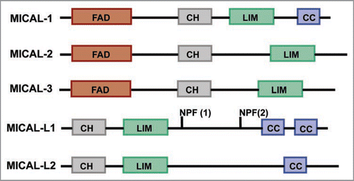
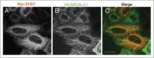
Figure 2 Co-localization of EHD1 and MICAL-L1 to tubular recycling endosomes. HeLa cells were co-transfected with Myc-tagged EHD1 (A) and HA-tagged MICAL-L1 (B). After 16 h, cells were fixed/permeabilized and incubated with anti-Myc, and anti-HA primary antibodies, and appropriate secondary antibodies before being mounted on cover-slides. Images were obtained from a Zeiss LSM 5 confocal microscope, using a 63x objective with a numerical aperture of 1.4. Bar, 10 µm.