Figures & data
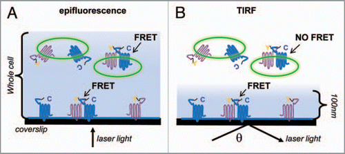
Figure 1 FRET in epifluorescence vs. TIRF. (A) A cell illuminated by direct epifluorescence. Y—YFP flourophore; C—CFP fluorophore. Shaded blue areas represent regions illuminated by laser light. In epifluorescence, fluorophores throughout the whole cell, including intracellular compartments, are illuminated. (B) The same cell illuminated under TIRF, where θ represents the critical angle. Only flourophores within ∼100 nm of the plasma membrane are illuminated.