Figures & data
Figure 1 Schematic diagram showing the domain organization of Npl3 homologues (A and B) and Gbp2 homologues (C and D) of Plasmodium falciparum and Homo sapiens. The protein names and species are written on the left hand side in each panel. The domain analysis was done using Scan Prosite at (expasy.org). (E) Superimposed image of the computer based structure model of the RR M domain of Plasmodium falciparum Gbp2 (shown in pink) on the RR M domain of Fir (2qfj) as a parent template (shown in yellow).
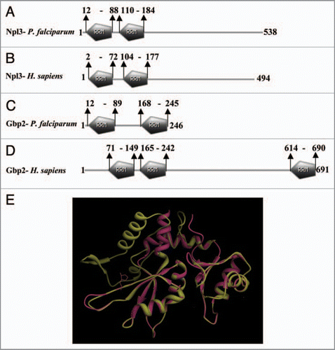
Figure 2 The computer based structure model of (A) PfU52 of Plasmodium falciparum and (B) Human UAP56 (1XTI). (C) The superimposed image.

Figure 3 (A) Comparison of the domain organization in the Ref proteins of Higher eukaryotes (i) and apicomplexans (ii). The conserved N and C terminal domains flanking the RR M motif are shown as RE F-N and RE F-C respectively. N-v and C-v represents the N and C terminal variable regions. (The figure has been prepared with the help of information given in ref. 55). (B) Schematic diagram showing the domain organization in the RE F family of (1) Plasmodium falciparum, (2) Plasmodium vivax, (3) Toxoplasma gondii, (4) Sacchromyces cerevisiae and (5) Homo sapiens. The domain analysis was done using Scan Prosite at (http://expasy.org). The protein names and species are written on the left hand side.
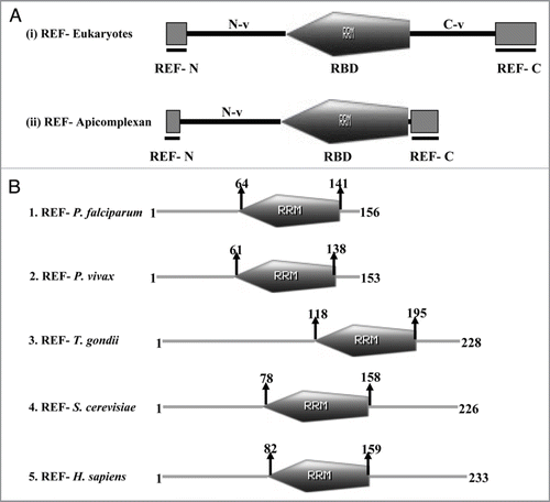
Figure 4 The computer based structure modeling of (A) PfRE F of Plasmodium falciparum based on the (B) 2E5 h-template (C) The superimposed image.
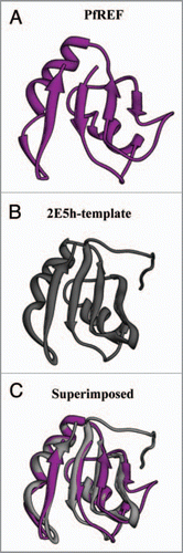
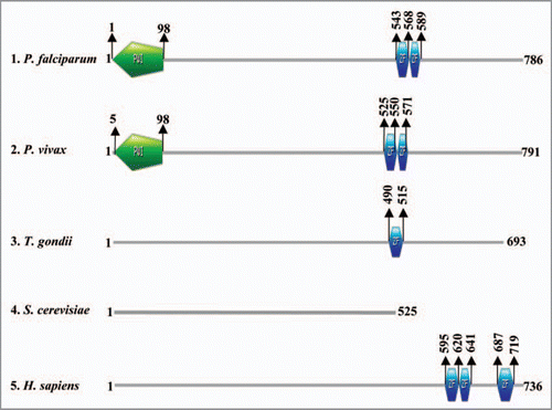
Figure 5 Schematic diagram showing the domain organization in various homologues of Nab2 in (1) Plasmodium falciparum, (2) Plasmodium vivax, (3) Toxoplasma gondii, (4) Sacchromyces cerevisiae and (5) Homo sapiens. The domain analysis was done using Scan Prosite at (http://expasy.org). The names of species are written on the left hand side.