Figures & data
Figure 1 TuMV replication factories are associated with ER and co-allign with microfilaments. Nicotiana benthamiana cells expressing mCherry-tagged TuMV-induced replication factories and ER -resident GFP (A) or the actin domain of fimbrin fused to GFP (B) observed by confocal microscopy at 4 days post-agroinfiltration. Photographs are a three-dimensional rendering of 40 1-µm thick slices that overlap by 0.5-µm. Scale bar, 10 µm.
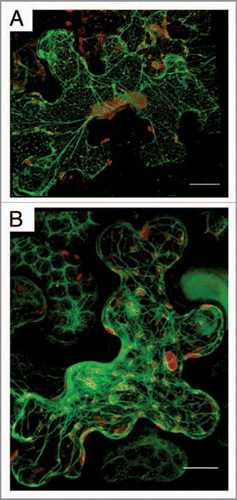
Figure 2 Model for the formation of virus-induced vesicles. The blue sphere represents the nucleus, while the brown structure the ER. Partially transparent virus-induced vesicles are in light blue. Green ribbons and red spheres and rods depict viral RN As and proteins, respectively. Host proteins are the orange cubes, and the brown and yellow structures are ribosomes.