Figures & data
Figure 1 Chemical structure of dehydroergosterol. Differences to ergosterol and cholesterol are indicated in red, while additional differences between DHE and cholesterol are indicated in green. Accordingly, DHE differs from ergosterol just in one additional double bound in the steroid backbone (red).
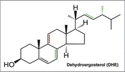
Figure 2 Bleach-rate-based image segmentation to visualize DHE in C. elegans. The figure illustrates the method of pixel-wise bleach-rate fitting used to distinguish probe fluorescence from autofluorescence in living nematodes. Various mathematical decay models, like a mono-exponential decay [mmo(t)] or stretched exponential decay [mst(t)] were fitted to the bleaching kinetics of the fluorescent sterol dehydroergosterol (DHE) in the nematode Caenorhabditis elegans in a pixel-wise manner. Images of DHE-stained worms were repeatedly acquired on a UV-sensitive wide field microscope to generate an input sequence. In every pixel position the intensity decay caused either by bleaching of DHE or of autofluorescence was fitted to the respective model, as exemplified for one pixel (white circle) in the diagram. Our software generates an amplitude map (upper green image) and a bleach rate map (lower image in FIRE LUT ).
![Figure 2 Bleach-rate-based image segmentation to visualize DHE in C. elegans. The figure illustrates the method of pixel-wise bleach-rate fitting used to distinguish probe fluorescence from autofluorescence in living nematodes. Various mathematical decay models, like a mono-exponential decay [mmo(t)] or stretched exponential decay [mst(t)] were fitted to the bleaching kinetics of the fluorescent sterol dehydroergosterol (DHE) in the nematode Caenorhabditis elegans in a pixel-wise manner. Images of DHE-stained worms were repeatedly acquired on a UV-sensitive wide field microscope to generate an input sequence. In every pixel position the intensity decay caused either by bleaching of DHE or of autofluorescence was fitted to the respective model, as exemplified for one pixel (white circle) in the diagram. Our software generates an amplitude map (upper green image) and a bleach rate map (lower image in FIRE LUT ).](/cms/asset/2941780d-d769-47bf-b086-6d76a856c116/kcib_a_10911972_f0002.gif)
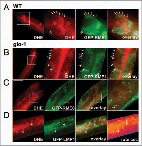
Figure 3 Co-localization of DHE with GFP-tagged markers of the endosomal pathway. N2 nematodes (A) or glo1-mutant worms (B-D) expressing either GFP-RME 1 (A and B); GFP-RME 8 (C) or GFP-LMP1 (D) were labeled with DHE by feeding a DHE-cyclodextrin solution as described.Citation7 DHE was visualized by repeated imaging on a UV-sensitive wide field microscope and calculation of the bleaching kinetics of the sterol versus autofluorescence. The calculated amplitude image of the rapidly bleaching fraction resembled DHE and is shown in the most left panels of (C and D). Alternatively, in case of specimen movement, the DHE distribution was inferred from subtracting the last from the first frame of the stack (left in A and B), as described previously (ref. Citation7). Both methods gave identical results on sterol distribution. DHE was found in GFP-RME -1-positive basolateral recycling endosomes (arrows in A and B) and additional in large round storage organelles lacking this endosomal marker. These organelles also lacked GFP-LMP1 (arrowheads in D), but were surrounded by GFP-RME 8 (arrows in C, and inset). Sterol-containing tissue could be identified from the rapid bleaching of DHE being represented by a high bleach rate constant (exemplified for worms expressing GFP-LMP1; most right in D). High and low rate constants are given in blue/violet and orange/yellow, respectively. Bar, 20 µm.