Figures & data
Figure 1 Cux genes control dendrite branching and synaptogenesis. Cux1 and Cux2 regulate neuronal differentiation and control intrinsic mechanisms of dendrite development, spine formation and synaptic function in upper layers in the cortex. Upper part: Dendritic parterns in WT and Cux2-/- pyramidal neurons of the upper layers. Lower parts: Downregulation of Xlr3b and Xlr4b gene expression by Cux proteins contributes to dendritic spine differentiation. Cux1 and Cux2 bind and regulate different regions in the Xlr4b locus. Miniature excitatory postsynaptic current (mEPSCs) from layer II and III pyramidal cells of Cux2-/- mice were reduced in amplitude and frequency.
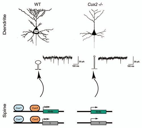
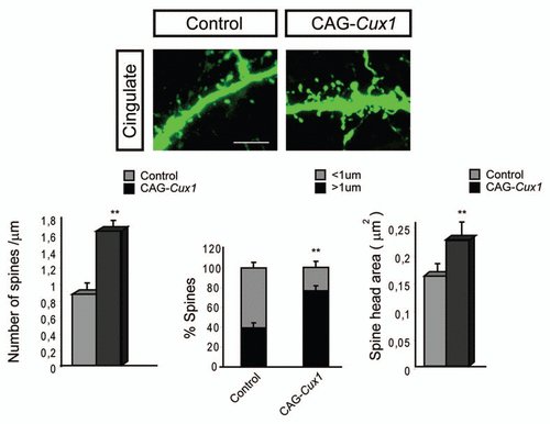
Figure 2 Dendritic spine formation in neurons of the cingulate cortex is stimulated upon Cux1 overexpression. Upper parts show representative confocal image of GFP positive spines in the cingulate cortex. These neurons had been electroporated with control or CAG-Cux1 plasmid. Scale bar represents 5 um. Lower parts show quantification of dendritic spine number, spine morphology and spine head area. Data in bar graphs depict mean ± SD. *p < 0.005, **p < 0.001, compared with control. This figure is a modification of Cubelos et al.Citation15