607
Views
22
CrossRef citations to date
0
Altmetric
Article Addendum

Ubiquitin receptor binding and signaling in primary human leukocytes

, , &
Pages 608-610 | Received 19 Aug 2020, Accepted 19 Aug 2010, Published online: 01 Nov 2010

Figures & data

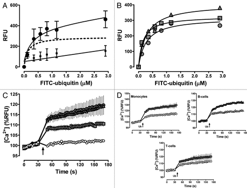
Figure 1 (A) FITC-ubiquitin binding to human monocytes (1 min, 4°C). Note that cells were centrifuged for 5 min to remove free FITC-ubiquitin in the cell culture supernatant. Data are mean ± Sem of duplicate measurements with monocytes from seven healthy blood donors. ●, FITC-ubiquitin binding; ■, non-specific binding, as assessed by binding of FITC-ubiquitin in the presence of 300 µm native ubiquitin; dashed line, specific binding curve (= total FITC-ubiquitin binding - non-specific binding; r2: 0.93). (B) Specific FITC-ubiquitin binding curves in monocytes (), B () and t cells () from a single blood donor, determined as in (A). (C) ubiquitin (3 µm) induced Ca2+ flux in monocytes (

), B-(
) and t-cells (
) from (B). Data are mean ± SEM from 10 cell cultures per cell type. Arrows indicate the time point when ubiquitin or vehicle (○) was added. (D) Inhibition of the ubiquitin induced Ca2+ signal by the PLC inhibitor U73122 (10 µM, open symbols). Data are mean ± SEM from three cell cultures per cell type. The weak PLC inhibitor U73343 (10 µM; grey symbols) was used as a negative control for U73122. Same cells as in B and C. Arrows indicate the time point when ubiquitin (3 µM) was added. RFU: relative fluorescence units.

Figure 1 (A) FITC-ubiquitin binding to human monocytes (1 min, 4°C). Note that cells were centrifuged for 5 min to remove free FITC-ubiquitin in the cell culture supernatant. Data are mean ± Sem of duplicate measurements with monocytes from seven healthy blood donors. ●, FITC-ubiquitin binding; ■, non-specific binding, as assessed by binding of FITC-ubiquitin in the presence of 300 µm native ubiquitin; dashed line, specific binding curve (= total FITC-ubiquitin binding - non-specific binding; r2: 0.93). (B) Specific FITC-ubiquitin binding curves in monocytes (), B () and t cells () from a single blood donor, determined as in (A). (C) ubiquitin (3 µm) induced Ca2+ flux in monocytes (Display full size), B-(Display full size) and t-cells (Display full size) from (B). Data are mean ± SEM from 10 cell cultures per cell type. Arrows indicate the time point when ubiquitin or vehicle (○) was added. (D) Inhibition of the ubiquitin induced Ca2+ signal by the PLC inhibitor U73122 (10 µM, open symbols). Data are mean ± SEM from three cell cultures per cell type. The weak PLC inhibitor U73343 (10 µM; grey symbols) was used as a negative control for U73122. Same cells as in B and C. Arrows indicate the time point when ubiquitin (3 µM) was added. RFU: relative fluorescence units.