Figures & data
Figure 1 Legionella pneumophila uptake and intracellular proliferation in the RacH-null mutant. (A) Bacterial uptake was measured by plating parental AX2 or RacH-null cells on 24-well tissue culture plates and adding L. pneumophila at a MOI of 10. To promote uptake, the bacteria were sedimented onto the cell layer by centrifugation. At the time indicated on the abscissa, extracellular bacteria were killed by short incubation with gentamycin and the number of L. pneumophila per cell was calculated by determining the colony forming units (CFU) per ml and normalizing for the cell number. (B) L. pneumophila intracellular growth in parental AX2 cells or RacH-null mutant was measured as above by using a MOI of 1 and by omitting gentamycin treatment. The CFU was determined at the time indicated in the abscissa. Under the conditions used, extracellular bacteria failed to grow.
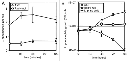
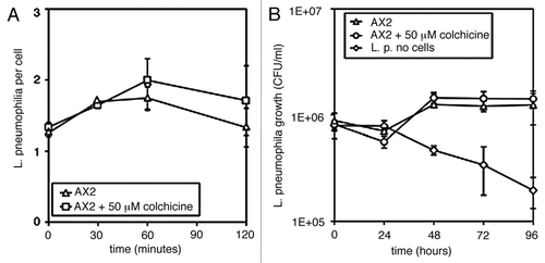
Figure 2 Legionella pneumophila uptake (A) and intracellular proliferation (B) in cells treated with the microtubule inhibitor colchicine. AX2 cells were preincubated on ice for 15 min, colchicine at 0.05 mM was added and the cells were warmed up to 25°C before addition of the bacteria. L. pneumophila uptake (A) and growth (B) were assessed as described in the legend to .