Figures & data
Figure 1 Dendritic spine structure and visual cortical function. (A) For each experimental group we reported the Immaturity index (gray) versus optical activation (yellow) both normalized to control values for comparison. Con, control; DR, dark-reared; DR2h, dark-reared then exposed to light for 2 h; DR2d, dark-reared then exposed to light for 2 days; DR7d, dark-reared then exposed to light for 7 days, 2hCPP, dark-reared injected with CPP and exposed to light for 2 h; DRdia, dark-reared injected with diazepam daily for 7 days. (B) Correlation analysis between immaturity index and cortical activation. For light re-exposure on the timescale of days, there is a high correlation (R2 = 0.91) between structural and functional properties, with the exception of DR2h time point (blue dot).
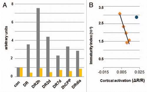
Figure 2 Visual experience does not alter the density of dendritic spines in primary visual cortex. The density of dendritic spines was determined by counting the number of spines per dendrite and dividing by the length of the dendrite in 3D. Density was calculated in the cortex of animals with different visual exposure. Here are reported the individual measurements per animal (asterisks) and the average per group (solid diamond). The density of spines was not different between different groups (Mann-Whitney; p > 0.05). N, number of animals.
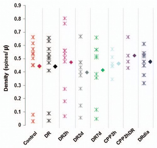
Figure 3 Visual experience controls the length of dendritic spines in primary visual cortex. The length of dendritic spines was determined by measuring each spine in the field of view in each condition. Empty triangles represent the average value per animal while solid triangles show the average value for the group. All deprived animal groups are significantly different from the controls (Mann-Whitney, p < 0.05). N, number of animals.
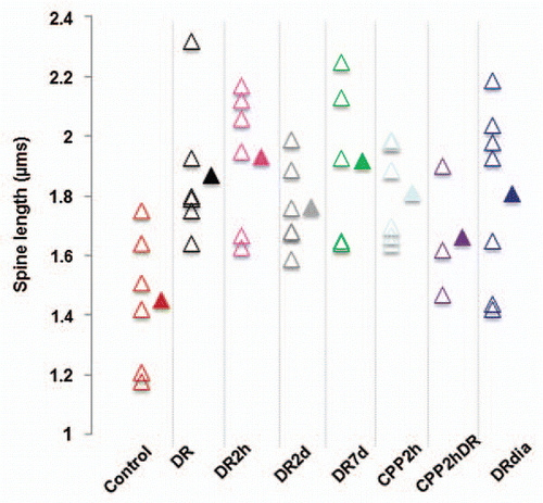
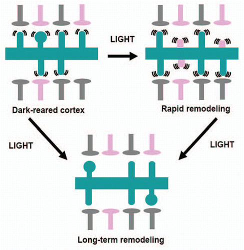
Figure 4 Light exposure induces a rapid remodeling of visual circuitry followed by stabilization. Two different phenomena modulate spine motility: one fast (within 2 h) and one slow (within 7 days). Slow recovery may be dependent on the initial fast response or may proceed through different mechanisms. Rapid changes in structural and functional events are mediated by NMDA receptors, while long-term changes are in part due to GABAergic transmission. Pink presynaptic terminals denote inappropriate connections. Gray terminals denote presynaptic terminal that form networks that serve visual processing. Pink spines denote inappropriate postsynaptic synapses in the processes of retraction.