372
Views
3
CrossRef citations to date
0
Altmetric
Mini Review

The Nucleolinus

A disappearing, forgotten, and (maybe) misnamed organelle

Pages 147-149 | Received 17 Dec 2010, Accepted 18 Dec 2010, Published online: 01 Mar 2011

Figures & data

Figure 1 Unactivated surf clam (Spisula solidissima) oocyte. A large tetraploid nucleus (germinal vesicle; GV) is present, within which lie a prominent nucleolus (arrow) and nucleolinus (arrowhead). The nuclear envelope begins to disintegrate within the first two minutes of activation and the GV is indistinguishable by 10–12 minutes post-fertilization. The nucleolus disappears within 5–6 minutes of fertilization, and the nucleolinus persists for 4–5 minutes by itself. Size bar = 15 µm.
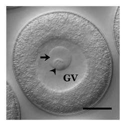
Figure 1 Unactivated surf clam (Spisula solidissima) oocyte. A large tetraploid nucleus (germinal vesicle; GV) is present, within which lie a prominent nucleolus (arrow) and nucleolinus (arrowhead). The nuclear envelope begins to disintegrate within the first two minutes of activation and the GV is indistinguishable by 10–12 minutes post-fertilization. The nucleolus disappears within 5–6 minutes of fertilization, and the nucleolinus persists for 4–5 minutes by itself. Size bar = 15 µm.

Figure 2 Distinguishing the nucleolinus from the nucleolus at the molecular level. The left-hand column of panels (A, D, G and J) are DIC images of oocytes. The middle column (B, E, H and K) shows the various probe signals in color brightfield (B) or immunofluorescence (E, H and K). The right hand column of panels (C, F, I and L) are overlays showing the specific labeling patterns against the DIC backgrounds. (A–C) in situ hybridization showing the localization of NLi-1 RNA to the nucleolinus (arrowhead) in an unactivated oocyte, and its absence from the nucleolus (arrow). Note that the nucleolus and nucleolinus swell slightly during the three-day in situ hybridization regimen. (D–F) localization of an antigen recognized by mcAb NLi-26 present in the nucleolus, but absent from the nucleolinus. (G–I) localization of an antigen recognized by mcAb NLi-9, which is absent from the nucleolus but localizes to a small zone near one pole of the nucleolinus in unactivated oocytes. After fertilization, the NLi-9 antigen expands across the nucleolinus (J–L). Oocytes shown in (D–L) are slightly compressed under the cover glass; the borders of the GV are therefore not as clearly distinguishable.
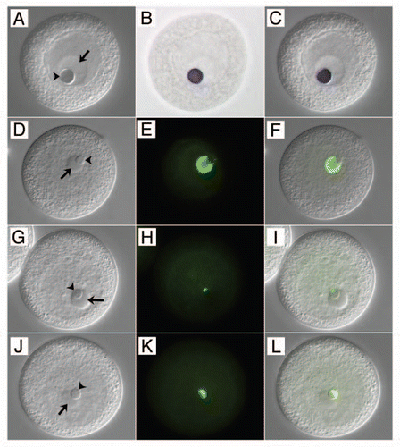
Figure 2 Distinguishing the nucleolinus from the nucleolus at the molecular level. The left-hand column of panels (A, D, G and J) are DIC images of oocytes. The middle column (B, E, H and K) shows the various probe signals in color brightfield (B) or immunofluorescence (E, H and K). The right hand column of panels (C, F, I and L) are overlays showing the specific labeling patterns against the DIC backgrounds. (A–C) in situ hybridization showing the localization of NLi-1 RNA to the nucleolinus (arrowhead) in an unactivated oocyte, and its absence from the nucleolus (arrow). Note that the nucleolus and nucleolinus swell slightly during the three-day in situ hybridization regimen. (D–F) localization of an antigen recognized by mcAb NLi-26 present in the nucleolus, but absent from the nucleolinus. (G–I) localization of an antigen recognized by mcAb NLi-9, which is absent from the nucleolus but localizes to a small zone near one pole of the nucleolinus in unactivated oocytes. After fertilization, the NLi-9 antigen expands across the nucleolinus (J–L). Oocytes shown in (D–L) are slightly compressed under the cover glass; the borders of the GV are therefore not as clearly distinguishable.