Figures & data
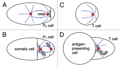
Figure 1 Schematic of centrosome positioning in Caenorhabditis elegans blastomeres and T lymphocytes. (A) single-cell stage of the worm embryo. (B) two-cell stage of the embryo. (C) isolated T lymphocyte, a white blood cell in suspension. (D) T cell conjugated with an antigen-presenting cell. Cell boundaries are in black, centrosomes in red and microtubules in blue. Gray arrows show translational, rotational and oscillatory movements.