Figures & data
Figure 1 A net-like structure is observed during fusion of HEK-TetOn cells with CHO cells. (A and B) HEK-TetOn cells that express FGFRL1 on their cell surface were seeded together with CHO cells that had been transfected with a GFP construct, which is under the control of the Tet transactivator protein. Photographs of two syncytial cells were taken at different magnifications. FGFRL1 at the surface of the syncytia was stained with a monoclonal antibody, followed by a Cy3 labeled secondary antibody (red). GFP (green) is expressed after the Tet transactivator protein has diffused from the HEK-TetOn cells to the CHO cells. At higher magnification, a net-like structure with pores (arrow) becomes visible. (C and D) The net-like structure is also observed when the HEK-TetOn cells were cultivated without the CHO cells. In this case, FGFRL1 was stained with the monoclonal antibody, followed by a Cy2-labeled secondary antibody (green). When the HEK-TetOn cells were seeded at higher density, the net-like structures are found to be distributed around the entire cells.
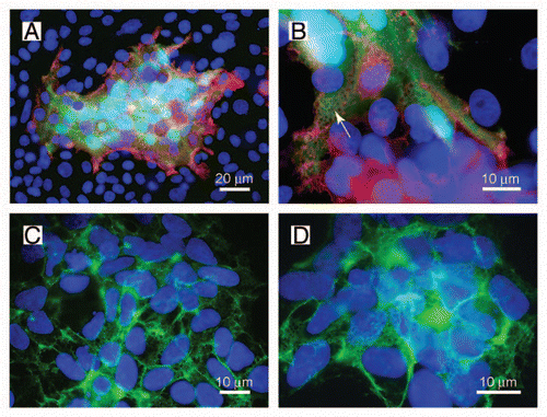
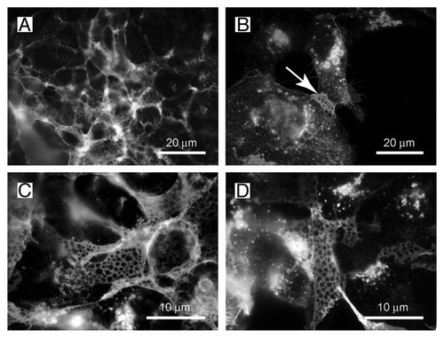
Figure 2 The net-like structures on the surface of HEK-TetOn cells show pores with a diameter of 1 ?m. (A and B) The net-like structures are preferentially observed at membrane regions where two cells touch each other (arrow). (C and D) The diameter of the pores is approximately 1 µm as determined at higher magnification.