Figures & data
Figure 1 Sebaceous gland size is significantly reduced by isotretinoin at 8 weeks of treatment compared to one week. Hematoxylin and eosin sections of skin biopsies were taken from patients before and at approximately 8 weeks or 1 week of isotretinoin treatment. (A) A total of 16 sections at baseline and 12 sections at 8 weeks of treatment were taken from 8 patients and analyzed using image analysis software. The mean area (±SEM) of sebaceous glands in the baseline samples was 0.17 ± 0.04 mm2 compared to 0.04 ± 0.07 mm2 in the 8-week samples; a four-fold reduction in size (p = 0.009). (B) A total of 17 sections at baseline and 19 sections at 1 week of treatment were taken from six patients and analyzed using image analysis software. In contrast to 8 weeks of isotretinoin treatment, the mean area (±SEM) of sebaceous glands in the 1-week baseline samples was 0.23 ± 0.09 mm2 compared to 0.12 ± 0.02 mm2 after 1 week of treatment, which was not significant using a paired t-test (p = 0.16). Representative images from three patients at each time point are shown at original magnification of 100x. Magnification bars = 250 µm. *Initial publication: Journal of Investigative Dermatology advance online publication, 6 November 2008; doi:10.1038/jid.2008.338.
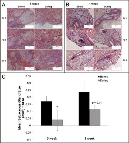
Figure 2 QPCR verifies the magnitude and direction of gene array changes. In order to verify the direction and magnitude of the changes in gene expression induced by isotretinoin in the gene array expression analysis, QPCR was performed using primers to select genes whose expression was significantly changed by 13-cis RA in the array analysis. Data represent the mean ± SEM of the fold-change in gene expression as determined by QPCR in four subjects compared to array analysis performed in eight subjects: 3β-hydroxysteroid dehydrogenase (3βHSD1), HMG CoA reductase (HMGCR), phospholipase A2 group 7(PLAG7), insulin induced gene 1 (INSIG), carnitine acyltransferase (CRAT) and zinc finger binding protein 145 (ZBTB16). Significant changes in gene expression with the microarray were identified using Significance Analysis of Microarrays (SAM) computer software and QPCR results were analyzed by the REST-XL software program; *p < 0.05 was considered significant for both analyses.
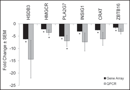
Figure 3 One week Clustering Diagram. Hierarchical clustering was used to compute a dendogram that assembled all samples and genes into a single tree. Normalized gene array expression data from each patient’s baseline and treatment biopsy was imported into dChip software version 1.3. Each row represents a single gene and each column represents a patient sample. B = baseline and A = after treatment. The color reflects the level of expression when compared to the mean level of expression for the entire 1 week biopsy set.
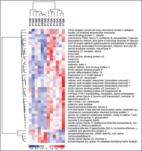
Figure 4 Temporal effects of isotretinoin.
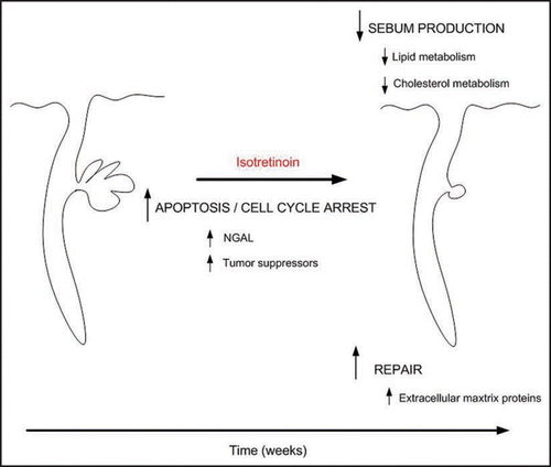
Table 1 8-week patient demographics
Table 2 Select genes changed significantly by isotretinoin in patient skin at 8 weeks
Table 3 Protein domains and pathways enriched within the genes significantly changed at 8 weeks
Table 4 Pathways enriched within the genes significantly changed at 8 weeks
Table 5 Functional categorization of significantly changed genes at 1 week isotretinoin
Table 6 Promoter analysis of genes significantly changed at 1 week of isotretinoin therapy