Figures & data
Figure 1. Determination of steroid hormone concentrations in blood and suction blister fluids. (A–F) Concentrations of DHEA, androstenedione and testosterone in blood and suction blister fluids of young (n = 8) and old male (n = 8) subjects were determined by UPLC-MS/MS after solid phase extraction. Blood concentrations of DHEA, androstenedione and testosterone are shown on the left side (A, C and E) and corresponding SBF concentrations on the right side (B, D and F). Statistical significance differences were marked with an asterisk (p < 0.05).
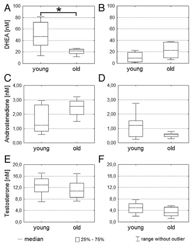
Figure 2. Steroid hormone concentrations in SBF from different skin sites. (A–C) SBF from upper back (n = 8) and forearm (n = 8) of young male subjects were generated in parallel. Concentrations of DHEA, androstenedione and testosterone were subsequently determined by UPLC-MS/MS after solid phase extraction. Statistical significance differences were marked with an asterisk (p < 0.05).
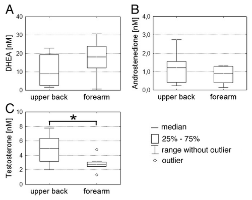
Table 1. Relative steroid hormone concentrations in cell culture supernatants after DHEA treatment
Figure 3. 3β-HSD type I expression in vitro. Confluent cultured SZ95 sebocytes (SZ95), primary dermal fibroblasts (HDF) and primary epidermal keratinocytes (NHEK) were harvested. Total protein fractions were isolated and subjected to western blot analysis using an anti 3β-HSD type I antibody. An anti-GAPDH antibody served as loading control.
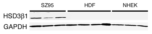
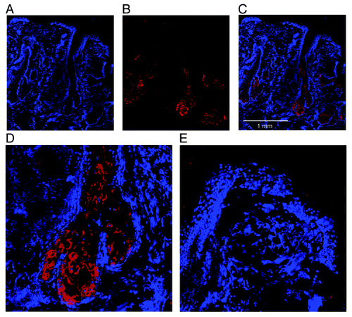
Figure 4. 3β-HSD type I expression in vivo. Skin biopsies of human scalp skin were cryosectioned. DAPI staining (A) was used to visualize histological structures. Positive 3β-HSD type I immunoreactivity was confirmed using an anti-3β-HSD type I antibody (B). A merge image (C) of DAPI staining and 3β-HSD type I immunoreactivity showed that the immunoreactivity was restricted to sebaceous glands and lower parts of the hair follicle duct, which is confirmed by a magnification of a sebaceous gland (D) and epidermis (E).