Figures & data
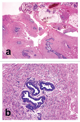
Figure 1 Histopathological findings. (A) Groups of proliferating and branching ductal structures embedded in a fibrous connective tissue stroma in the subcutaneous fat tissue (haematoxylin and eosin, original magnification x6.25). (B) Dilated ducts with intraluminal secretion are lined by cuboidal cells. The connective tissue stroma was sparse around the ducts (haematoxylin and eosin, original magnification x100).