Figures & data
Figure 1. Schematic display of the three stress axis and their activation by a wide variety of stressors as well as selected key effects on the immune system and subsequently chronic disease and inflammaging regulated by inflammatory processes. 1sympathetic nervous system; 2hypothalamus pituitary adrenal axis; 3neurotrophin neuropeptide axis.
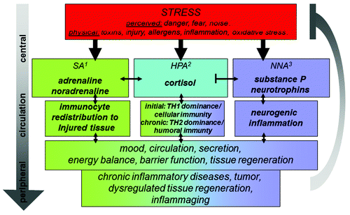
Figure 2. Schematic drawing of the splenic white pulp as the screening area for peripheral inflammation. In the periphery an antigen is taken up by an antigen presenting cell (APC). Cytokines are produced and reaches the circulation. The APC re-migrates into the spleen via the central artery. From here the blood enters the marginal sinus (MS) via the central arterioles (CA) and through open cavities between the endothelial cells surrounding the periarteriolar lymphoid sheaths (PALS) and marginal zone (MZ) circulating cells and effector molecules can enter the splenic white pulp (PALS+MZ). The antigen is presented to lymphocytes within a special microenvironment generated by the effector molecules and the proximity to local nerve fibers which are activated upon stress. The neuro-immune crosstalk itself governs local neurotransmitter- and neuropeptide release and thereby tunes the TH1/TH2 cytokine balance. Vice versa those effector molecules as well as local produced neurotransmitters and neuropeptides finally abandon the spleen via the splenic vein and re-circulate with potential effects on peripheral inflammation and potentially inflammaging.
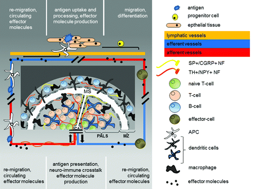
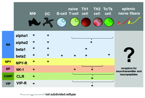
Figure 3. Distribution of neurotransmitter and neuropeptide receptors on immune cells. The picture gives an overview about the distribution of neurotransmitter- and neuropeptide receptors on immune cells. The question mark indicates a missing link in neuro-immune crosstalk, the distribution of cytokine receptor on splenic nerve fibers. MΦ, Macrophage; DC, dendritic cell; Tc, T-cytotoxic cell; Ts, T-supressor cell; NA, noradrenalin; NPY, neuropeptide Y; SP, substance P; CGRP, calcitonine-gene related peptide; VIP, vasointestinal peptide.