Figures & data
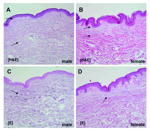
Figure 1. A comparison between male (A and C) and female (B and D) endogenous aged skin. Staining via hematoxylin eosin (A and B) and elastica staining (C and D), respectively, revealed that the dermis in the male is significantly thicker than in the female. In contrast, epidermis and subcutaneous tissue are significantly thicker in the female.