573
Views
8
CrossRef citations to date
0
Altmetric
Report

Auto/paracrine nicotinergic peptides participate in cutaneous stress response to wounding

, , &
Pages 324-330 | Received 27 Aug 2012, Accepted 19 Oct 2012, Published online: 01 Jul 2012

Figures & data

Figure 1. rSLURP-1 and -2 exhibit opposite effects on lateral migration of human KCs under agarose. Normal human KCs were seeded in the 3 mm wells of the AGKOS plates measuring random cell migration, exposed to increasing concentrations of rSLURP-1 (-●-) or -2 (-■-) dissolved in growth medium, and incubated for 10 d with daily refreshing the media. Migration distance was measured as described in Materials and Methods. Migration distance of control KCs incubated in growth medium without rSLURPs was taken as 100%, and the results expressed as percent of intact controls. Asterisks = p < 0.05 compared with control.
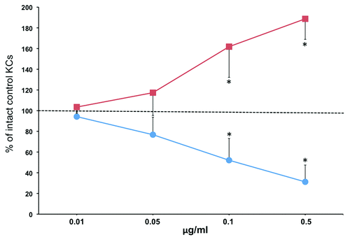
Figure 1. rSLURP-1 and -2 exhibit opposite effects on lateral migration of human KCs under agarose. Normal human KCs were seeded in the 3 mm wells of the AGKOS plates measuring random cell migration, exposed to increasing concentrations of rSLURP-1 (-●-) or -2 (-■-) dissolved in growth medium, and incubated for 10 d with daily refreshing the media. Migration distance was measured as described in Materials and Methods. Migration distance of control KCs incubated in growth medium without rSLURPs was taken as 100%, and the results expressed as percent of intact controls. Asterisks = p < 0.05 compared with control.

Figure 2. Anti-SLURP antibodies interfere with wound epithelialization. The excisional wounds in the skin of BALB/c mice were treated daily with microinjections of 10 μg/ml of anti-SLURP-1 or -2 antibodies or their mixture vs. 10 μg/ml of NIgG vs. plain saline (control) for the periods of time indicated in the graph. The wound epithelialization rate was measured as detailed in Materials and Methods. The percentage of epidermal closure was calculated as a ratio of the epithelialized to the initial wound area. Circles = p < 0.05 compared with control.
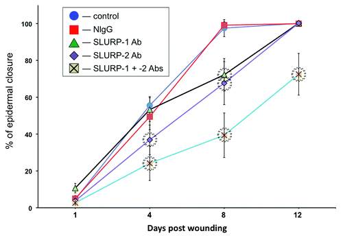
Figure 2. Anti-SLURP antibodies interfere with wound epithelialization. The excisional wounds in the skin of BALB/c mice were treated daily with microinjections of 10 μg/ml of anti-SLURP-1 or -2 antibodies or their mixture vs. 10 μg/ml of NIgG vs. plain saline (control) for the periods of time indicated in the graph. The wound epithelialization rate was measured as detailed in Materials and Methods. The percentage of epidermal closure was calculated as a ratio of the epithelialized to the initial wound area. Circles = p < 0.05 compared with control.

Figure 3. Anti-SLURP antibodies increase inflammation of cutaneous wounds. The murine cutaneous wounds treated as described in the legend to were harvested, homogenized and used in the ELISA assays of the biomarkers of inflammation MPO, IL-1β. IL-6 and TNFα. Circles = p < 0.05 compared with control.
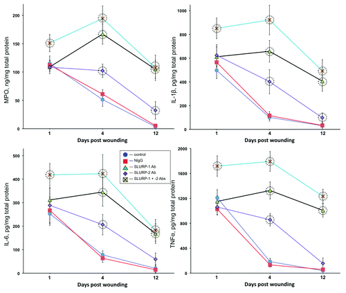
Figure 3. Anti-SLURP antibodies increase inflammation of cutaneous wounds. The murine cutaneous wounds treated as described in the legend to Figure 2 were harvested, homogenized and used in the ELISA assays of the biomarkers of inflammation MPO, IL-1β. IL-6 and TNFα. Circles = p < 0.05 compared with control.