Figures & data
Figure 1. BPA from the mother’s exposure can pass through the placenta and expose the developing fetus. Maternal 17β-estradiol, on the other hand, is metabolized in the placenta and does not reach the fetus. Therefore, the levels of estrogenic active BPA can readily exceed endogenous levels of 17β-estradiol in the developing fetus, also at low-level exposure.
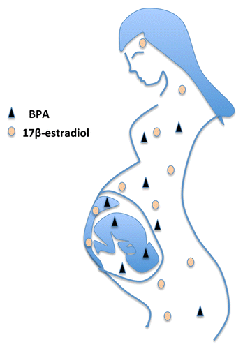
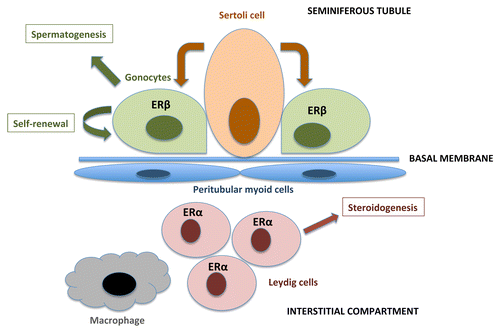
Figure 2. A schematic view of the developing testis: cells and functions, including expressions of the ERs. Sertoli cells in the seminiferous tubules comprise a stem cell niche and support the gonocytes (germ cells). The gonocytes express ERβ during fetal and early neonatal development, and are capable of differentiation into spermatids through several stages of spermatogonia/spermatocytes. Leydig cells are located in the interstitial compartment, they express ERα and are responsible for hormone synthesis.