Figures & data
Figure 1. Schematic representation of the FSHD locus in control individuals, patients with contraction dependent (FSHD1) and patients with contraction independent (FSHD2) FSHD. D4Z4 repeat units are indicated with triangles, the additional DUX4 exon with stabilizing polyA signal distal to the repeat is indicated with a box. Common chromatin features of D4Z4 in FSHD1 and FSHD2 are decreased CpG methylation levels and a more relaxed chromatin structure facilitating the expression of DUX4 from the telomeric repeat unit. Overall reduction in CpG methylation of D4Z4 in FSHD is schematically indicated with reduced number of lollipop symbols
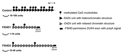
Figure 2. Scatterplot of ChCS in fibroblasts (A) or myoblasts (B) of controls, FSHD1 and FSHD2 patients. Individual samples are represented by individual data points. Mean values with 1xSD are indicated.
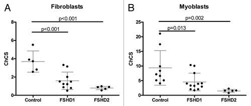
Table 1. Summary of analyses of age corrected CSS and ChCS
Figure 3. Scatterplot of ChCS vs. age-corrected CSS in fibroblast (A) and in myoblast samples (B). Male samples are represented by closed triangles, female samples by open triangles.
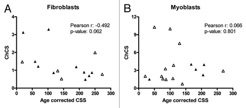
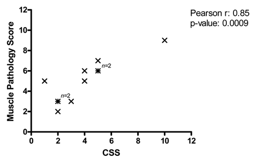
Figure 4. Relationship analysis between the CSS and MPS. There is a significant correlation between MPS and CSS (p = 0.0009). Note that the analysis was done without age correction.