Figures & data
Figure 1. BRCA1 promoter CpG island hypermethylation is associated with transcriptional silencing. (A) Pyrosequencing analysis of BRCA1 CpG island demonstrates hypermethylation in UACC-3199 and HCC-38 cancer cells. (B) Bisulfite genomic sequencing of eight individual clones in the BRCA1 promoter CpG island: examples of a normal breast and the breast cancer cell line HCC-38 are shown. Presence of a methylated or unmethylated cytosine is indicated by a black or white square, respectively. Black arrows indicate the position of the bisulfite genomic sequencing primers. (C) Real-time PCR expression of the BRCA1 transcript. (D) BRCA1 expression was also determined by western blot and the β-actin protein was used as a loading control. The UACC3199 and HCC-38 breast cancer cells show a hypermethylated CpG island in association with the downregulation of the BRCA1 protein. MDA-MB-231 (wild-type) and MDA-MB-436 (mutant) are shown as positive and negative controls for BRCA1 expression.
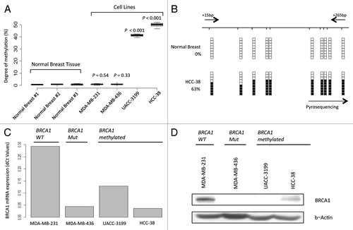
Figure 2. BRCA1 epigenetic inactivation predicts sensitivity to platinum-based chemotherapy. (A) Cell viability assessed by the MTT assays demonstrates that methylated (UACC3199 and HCC-38) and mutant (MDA-MB-436) BRCA1 cells both exhibit enhanced sensitivity to cisplatin and carboplatin in comparison with wild type and unmethylated MDA-MB-231 breast cancer cells. The corresponding IC50 values are shown. (B) Representative comet assays show DNA damage upon cisplatin use in the BRCA1 methylated or mutated cell lines. (C) Quantification of the obtained values from the comet assay. (E) BRCA1-hypermethylated cells are not able to repair DNA damage when cisplatin is used. The values of comet assays shown in box-plots demonstrate that both methylated and mutant BRCA1 cells experience permanent DNA damage when cisplatin is used that it is not observed in BRCA1 wild type or unmethylated cells (MDA-MB-231). (D) Relative changes in tumor size of UACC3199 (BRCA1 hypermethylated) and MDA-MB-231 (BRCA1 unmethylated) cancer cells xenografted in nude mice upon cisplatin use. Values shown at 28 d after the start of the chemotherapy treatment.
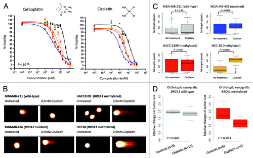
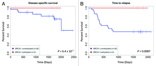
Figure 3. BRCA1 hypermethylation proves to be a predictor of good response to chemotherapy with cisplatin in ovarian cancer patients. (A) BRCA1 hypermethylation in patients with ovarian cancer is associated with longer time to relapse. (B) BRCA1 hypermethylation in patients with ovarian cancer is associated with improved disease-specific survival.