Figures & data
Figure 1. Unsupervised hierarchical clustering (A) and three-dimensional principal component analysis (PCA) of DNA methylation data (B) comparing samples belonging to subset #1, subset #2 and subset #4. The S refers to the subset number while the following number refers to the specific case as detailed in Table S1.
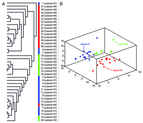
Figure 2. Supervised hierarchical clustering of differentially methylated genes (methylation index geometric mean difference > 0.35, p < 0.05). Comparisons were made between subsets #1 and #4 (A), subsets #1 and #2 (B) and subsets #2 and #4 (C). A gradient color scale represents the methylation index values; green as unmethylated and red as methylated.
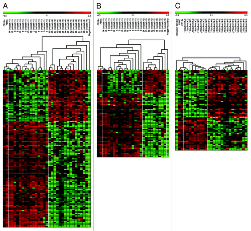
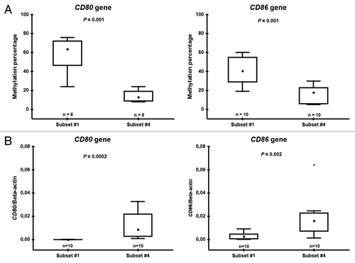
Figure 3. Pyrosequencing (A) and RQ-PCR (B) data on CD80 and CD86 in subset #1 and #4 patient samples. Boxes indicate the interquartile range (25–75%) while the small inner square indicates the median value. The whiskers represent the minimum and maximum values, except for outliers (circles) and extremes (stars).