637
Views
17
CrossRef citations to date
0
Altmetric
Research Paper

Zebularine partially reverses GST methylation in prostate cancer cells and restores sensitivity to the DNA minor groove binder brostallicin

, , , &
Pages 656-665 | Received 12 Feb 2013, Accepted 03 May 2013, Published online: 14 Jun 2013

Figures & data

Figure 1. (A) Cytotoxicity induced by increasing concentrations of brostallicin treatment in LNCaP (◻) and DU145 (●) cells. Results are reported as the percentage of inhibition relative to controls and are the mean ± SD of at least six replicates. °p < 0.05, *p < 0.01 vs LNCaP (B) GST total activity (left panel) and GST-pi protein expression (right panel) in LNCaP and DU145 cells. Actin is used as loading control in western blotting experiments. *p < 0.01 vs LNCaP (C) Pyrosequencing results showing the average of methylation percentage of 14 different CpG islands (corresponding with Y in the sequence at the bottom of the graph) in the GSTP1 promoter sequence of LNCaP (gray bars) and DU145 (white bars) cells. Results are expressed as mean ± SD of three independent evaluations. (D) GST activity (left panel) and GST-pi protein expression (right panel) in parental LNCaP and in two clones transfected with the human GSTP1 gene. Actin is used as loading control in western blotting experiments. *p < 0.01 vs LNCaP wt (E) Activity of brostallicin in parental LNCaP cells (●) and in LNCaP-derived clones 41 (■) and 54 (◻) overexpressing GST-pi. °p < 0.05, *p < 0.01 vs wt
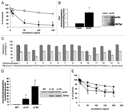
Figure 1. (A) Cytotoxicity induced by increasing concentrations of brostallicin treatment in LNCaP (◻) and DU145 (●) cells. Results are reported as the percentage of inhibition relative to controls and are the mean ± SD of at least six replicates. °p < 0.05, *p < 0.01 vs LNCaP (B) GST total activity (left panel) and GST-pi protein expression (right panel) in LNCaP and DU145 cells. Actin is used as loading control in western blotting experiments. *p < 0.01 vs LNCaP (C) Pyrosequencing results showing the average of methylation percentage of 14 different CpG islands (corresponding with Y in the sequence at the bottom of the graph) in the GSTP1 promoter sequence of LNCaP (gray bars) and DU145 (white bars) cells. Results are expressed as mean ± SD of three independent evaluations. (D) GST activity (left panel) and GST-pi protein expression (right panel) in parental LNCaP and in two clones transfected with the human GSTP1 gene. Actin is used as loading control in western blotting experiments. *p < 0.01 vs LNCaP wt (E) Activity of brostallicin in parental LNCaP cells (●) and in LNCaP-derived clones 41 (■) and 54 (◻) overexpressing GST-pi. °p < 0.05, *p < 0.01 vs wt

Figure 2. (A) Activity of brostallicin in the absence (left) or presence (right) of 0.05 μM 5-aza dC. Cells were treated with different concentrations of brostallicin and colonies formed stained with comassie blue. (B) Effect of 0.05 μM 5-aza dC on the growth of LNCaP cells. Black bar, controls, gray bar, 5-aza dC treated cells. Results are reported as mean ± SD of at least six replicates. *p < 0.01 vs wt (C) GST activity in untreated (black bar) or 0.05 μM 5-aza dC-treated (gray bar) LNCaP cells. Results are the mean ± SD of at three replicates. *p < 0.01 vs wt
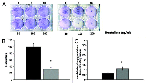
Figure 2. (A) Activity of brostallicin in the absence (left) or presence (right) of 0.05 μM 5-aza dC. Cells were treated with different concentrations of brostallicin and colonies formed stained with comassie blue. (B) Effect of 0.05 μM 5-aza dC on the growth of LNCaP cells. Black bar, controls, gray bar, 5-aza dC treated cells. Results are reported as mean ± SD of at least six replicates. *p < 0.01 vs wt (C) GST activity in untreated (black bar) or 0.05 μM 5-aza dC-treated (gray bar) LNCaP cells. Results are the mean ± SD of at three replicates. *p < 0.01 vs wt

Figure 3. (A) Cytotoxicity induced by increasing concentrations of brostallicin treatment in LNCaP cells in the absence (●) or in the presence of 100 (■) or 125 μM (◻) of zebularine. Results are reported as the percentage of inhibition relative to controls and are the mean ± SD of at least six replicates. *p < 0.01 vs cells not pretreated with zebularine. (B) GST activity in LNCaP cells treated with different concentrations of zebularine. Results are the mean ±− SD of at three replicates. *p < 0.01 vs cells not pretreated with zebularine. (C) Methylation specific–PCR in the GSTP1 (upper panel) and GSTM1 (lower panel) gene performed in LNCaP cells treated with zebularine. Lane 1 MW marker; Lanes 2 and 3 control cells methylated (M) and unmethylated (U); Lanes 4 and 5 100 μM zebularine treated cells, methylated (M) and unmethylated (U); Lanes 6 and 7 125 μM zebularine treated cells, methylated (M) and unmethylated (U); Lanes 8 and 9 internal controls (blanks) (D) Real time RT PCR performed in LNCaP cells untreated or treated with 100 or 125 mM zebularine. The table reports the expression ratio between treated and control cells (arbitrarily set to 1). The values haves been normalized for cyclophillin used as internal control.
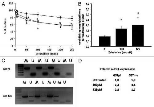
Figure 3. (A) Cytotoxicity induced by increasing concentrations of brostallicin treatment in LNCaP cells in the absence (●) or in the presence of 100 (■) or 125 μM (◻) of zebularine. Results are reported as the percentage of inhibition relative to controls and are the mean ± SD of at least six replicates. *p < 0.01 vs cells not pretreated with zebularine. (B) GST activity in LNCaP cells treated with different concentrations of zebularine. Results are the mean ±− SD of at three replicates. *p < 0.01 vs cells not pretreated with zebularine. (C) Methylation specific–PCR in the GSTP1 (upper panel) and GSTM1 (lower panel) gene performed in LNCaP cells treated with zebularine. Lane 1 MW marker; Lanes 2 and 3 control cells methylated (M) and unmethylated (U); Lanes 4 and 5 100 μM zebularine treated cells, methylated (M) and unmethylated (U); Lanes 6 and 7 125 μM zebularine treated cells, methylated (M) and unmethylated (U); Lanes 8 and 9 internal controls (blanks) (D) Real time RT PCR performed in LNCaP cells untreated or treated with 100 or 125 mM zebularine. The table reports the expression ratio between treated and control cells (arbitrarily set to 1). The values haves been normalized for cyclophillin used as internal control.

Figure 4. (A) Activity of brostallicin in the absence (●) or in the presence (◻) of 100 μM zebularine in DU145 cells. Results are reported as the percentage of inhibition relative to controls and are the mean ± SD of at least six replicates. (B) Activity of brostallicin in LNCaP-GST clone (clone 41) in the absence (●) or in the presence (◻) of 100 μM zebularine. Results are reported as the percentage of inhibition relative to controls and are the mean ± SD of at least six replicates. °p < 0.05 vs cells not pretreated with zebularine.
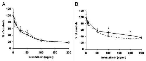
Figure 4. (A) Activity of brostallicin in the absence (●) or in the presence (◻) of 100 μM zebularine in DU145 cells. Results are reported as the percentage of inhibition relative to controls and are the mean ± SD of at least six replicates. (B) Activity of brostallicin in LNCaP-GST clone (clone 41) in the absence (●) or in the presence (◻) of 100 μM zebularine. Results are reported as the percentage of inhibition relative to controls and are the mean ± SD of at least six replicates. °p < 0.05 vs cells not pretreated with zebularine.

Figure 5. (A) Scheme of the treatment schedules used for the in vivo experiments performed in LNCaP-GST (left panel) and in LNCaP wt (right panel) xenografts. (B) In vivo antitumor activity of brostallicin in LNCaP-GST clone 41 xenografts. Mice were treated with brostallicin 0.4 mg/kg i.v. twice on days 6 and 20 after randomization (▲). Untreated mice (black diamond) °p < 0.05, 2 d after the last treatment with brostallicin. (C) In vivo antitumor activity of brostallicin in parental LNCaP wt xenografts. Mice were treated with brostallicin 0.4mg/kg i.v. twice on days 6 and 20 after randomization (●), zebularine 500 mg/kg daily ip twice for 7 d (■), first treatment: day 0 until day 6; second treatment: from day14 until day 20, or with the combination of zebularine and brostallicin (▲). Untreated mice (black diamond). *p < 0.01, combination group vs control and brostallicin groups and °p < 0.05 vs zebularine group. The tables report parameters derived from the curves in the two experimental settings and described at the bottom of the figures and in Materials and Methods.
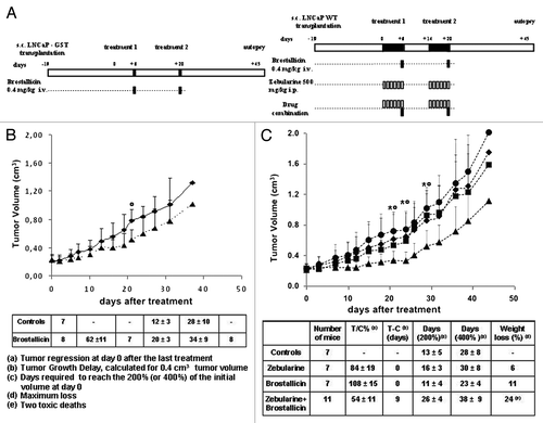
Figure 5. (A) Scheme of the treatment schedules used for the in vivo experiments performed in LNCaP-GST (left panel) and in LNCaP wt (right panel) xenografts. (B) In vivo antitumor activity of brostallicin in LNCaP-GST clone 41 xenografts. Mice were treated with brostallicin 0.4 mg/kg i.v. twice on days 6 and 20 after randomization (▲). Untreated mice (black diamond) °p < 0.05, 2 d after the last treatment with brostallicin. (C) In vivo antitumor activity of brostallicin in parental LNCaP wt xenografts. Mice were treated with brostallicin 0.4mg/kg i.v. twice on days 6 and 20 after randomization (●), zebularine 500 mg/kg daily ip twice for 7 d (■), first treatment: day 0 until day 6; second treatment: from day14 until day 20, or with the combination of zebularine and brostallicin (▲). Untreated mice (black diamond). *p < 0.01, combination group vs control and brostallicin groups and °p < 0.05 vs zebularine group. The tables report parameters derived from the curves in the two experimental settings and described at the bottom of the figures and in Materials and Methods.

Figure 6. (A) Immunohistochemistry analysis of GST-pi expression in parental LNCaP cells untreated (LNCaPwt) or pretreated for 7 consecutive days with zebularine 500mg/Kg (Zebularine). Positive control for GST expression is represented by untreated LNCaP-GST clone 41 growing in immunodeficient mice (LNCaP-GST). The first two panels report H&E staining. (B) Predicted CpG islands in GSTP1 gene according to MethPrimer program. (C) Methylation specific–PCR in the GSTP1 gene performed in LNCaP tumors obtained from untreated or zebularine treated mice (tumors taken after the last treatment and at autopsy) as reported in the figure. Calponin is used as internal control. The three last lanes represent the controls of methylation specific PCRs obtained by using unmethylated DNA (0), totally methylated DNA (100) or an equal mixture of them (50). (D) Real Time RT PCR performed in LNCaP tumors untreated or treated with zebularine. The table reports the cycle in which amplification occurs for GSTP1 and for cyclophillin used as internal control. The Delta Ct values calculated from the data are reported in the last column.
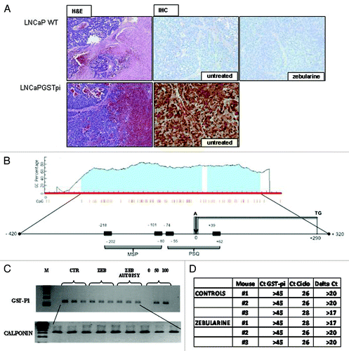
Figure 6. (A) Immunohistochemistry analysis of GST-pi expression in parental LNCaP cells untreated (LNCaPwt) or pretreated for 7 consecutive days with zebularine 500mg/Kg (Zebularine). Positive control for GST expression is represented by untreated LNCaP-GST clone 41 growing in immunodeficient mice (LNCaP-GST). The first two panels report H&E staining. (B) Predicted CpG islands in GSTP1 gene according to MethPrimer program. (C) Methylation specific–PCR in the GSTP1 gene performed in LNCaP tumors obtained from untreated or zebularine treated mice (tumors taken after the last treatment and at autopsy) as reported in the figure. Calponin is used as internal control. The three last lanes represent the controls of methylation specific PCRs obtained by using unmethylated DNA (0), totally methylated DNA (100) or an equal mixture of them (50). (D) Real Time RT PCR performed in LNCaP tumors untreated or treated with zebularine. The table reports the cycle in which amplification occurs for GSTP1 and for cyclophillin used as internal control. The Delta Ct values calculated from the data are reported in the last column.
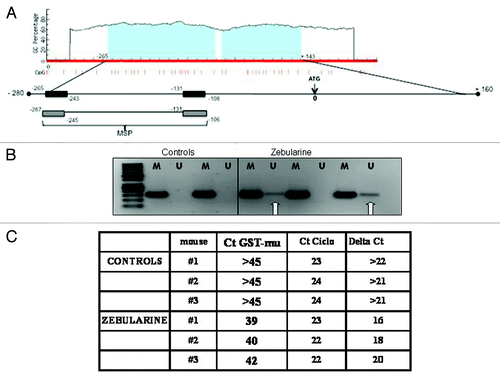
Figure 7. (A) Predicted CpG islands in GSTM1 gene according to MethPrimer program. (B) GSTM1 gene methylation specific–PCR performed in LNCaP tumors obtained from untreated or zebularine treated mice (tumors taken after the last treatment have been analyzed). M refers to methylated band and U to unmethylated amplified DNA. (C) Real Time RT PCR performed in LNCaP tumors untreated or treated with zebularine. The table reports the cycle in which amplification occurs for GSTM1 and for cyclophillin used as internal control. The Delta Ct values calculated from the data are reported in the last column.

Figure 7. (A) Predicted CpG islands in GSTM1 gene according to MethPrimer program. (B) GSTM1 gene methylation specific–PCR performed in LNCaP tumors obtained from untreated or zebularine treated mice (tumors taken after the last treatment have been analyzed). M refers to methylated band and U to unmethylated amplified DNA. (C) Real Time RT PCR performed in LNCaP tumors untreated or treated with zebularine. The table reports the cycle in which amplification occurs for GSTM1 and for cyclophillin used as internal control. The Delta Ct values calculated from the data are reported in the last column.
Supplemental material

Additional material

Download Zip (93.1 KB)

Reprints and Corporate Permissions

Please note: Selecting permissions does not provide access to the full text of the article, please see our help page How do I view content?

To request a reprint or corporate permissions for this article, please click on the relevant link below:

Academic Permissions

Please note: Selecting permissions does not provide access to the full text of the article, please see our help page How do I view content?

Obtain permissions instantly via Rightslink by clicking on the button below:

If you are unable to obtain permissions via Rightslink, please complete and submit this Permissions form. For more information, please visit our Permissions help page.