Figures & data
Figure 1. Re-expression of ARHI induces dormancy, and the downregulation of ARHI permits recurrent growth in cultures of ovarian cancer cells that are stimulated with VEGF. (A) Clonogenic growth of SKOv3-ARHI and Hey-ARHI cells after different treatments. The cells were divided into six groups: the blank control group (medium only), dormancy control group (cells treated with 1 μg/ml DOX for 14 d), VEGF control group (cells treated with 20 ng/ml VEGF for 14 d), dormancy group (cells treated with both 20 ng/ml VEGF and 1 μg/ml DOX for 14 d), recurrence group (cells treated with VEGF for 14 d and DOX for only 6 d followed by DOX removal), and recurrence control group (cells treated with 1 μg/ml DOX for 6 d followed by DOX removal). (B) Colony numbers of the SKOv3-ARHI (left) and Hey-ARHI (right) cell lines in the six groups were counted. The experiments were performed in triplicate, and the data are presented as the mean ± SD of the three separate experiments. **Compared with the VEGF control group, P < 0.01. ##Compared with the dormancy group, P < 0.01
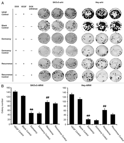
Figure 2. ARHI induces autophagy in ovarian cancer cells. (A) Expression of ARHI in the ovarian cancer dormancy-recurrence in-vitro model as measured by real-time PCR. Top: SKOv3-ARHI; bottom: Hey-ARHI. (B) Increased cleavage of LC3-I to LC3-II and higher ARHI expression were shown in the dormancy group, whereas the withdrawal of DOX resulted in decreased LC3-II and ARHI expression in SKOv3-ARHI (C) The percentage of autophagic cells was measured by acridine orange staining and flow cytometry. FL3H detects red fluorescence (650 nm), and FL1H detects blue fluorescence (488 nm). Dot plots that show increased autophage formation in the dormancy (VEGF+DOX treatment) group and decreased autophage formation after the transition of dormancy to recurrence (VEGF + DOX withdrawn). *Compared with the VEGF control group, P < 0.05, **Compared with the VEGF control group, P < 0.01; #Compared with the dormancy group, P < 0.01, ##Compared with the dormancy group, P < 0.01.
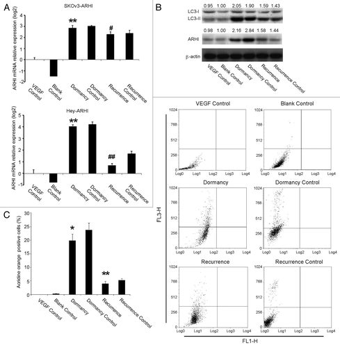
Figure 3. Anti-angiogenic genes are activated in dormant and repressed in recurrent ovarian cancer cells. (A) The expression of angiogenesis-related factors in dormancy and the recurrence transition measured by real-time PCR in SKOv3-ARHI cells. Of the 15 genes, the six genes (Ang1, Ang2, Ang4, TSP1, CDH1, and TIMP3) that were upregulated during dormancy and decreased again in recurrence were presented. (B) The expression of the six genes in the Hey-ARHI cells (C) Dynamics of TIMP3 and CDH1 expression in SKOv-3 ARHI cells. This expression was measured by real-time RT-PCR at four time points (0, 6, 10, and 14 d). (D) Dynamics of TIMP3 and CDH1 expression in Hey-3 ARHI cells. **Compared with the control group, P < 0.01. ##Compared with the dormancy group, P < 0.01
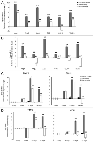
Figure 4. TIMP3 and CDH1 protein expression increases in dormancy, decreases in recurrence and are upregulated by 5-Aza-dC and TSA. The cells were divided into nine groups: the Blank control, VEGF control, Dormancy, Dormancy control, Recurrence and Recurrence control, which were mentioned in ; the 5-Aza-dC group: recurrence group treated with 5-Aza-dC for the last 72 h; the TSA group: recurrence group treated for TSA for the last 24 h; the 5-Aza-dC+TSA group: recurrence group treated with 5-Aza-dC for the last 72 h and with TSA for the last 24 h. The cells were stained with CDH1 and TIMP3 antibodies with the indicated concentrations. Brown staining means positive.
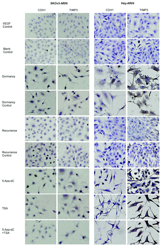
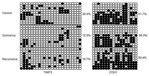
Figure 5. DNA methylation of TIMP3 and CDH1 decreases during dormancy and increases during recurrent growth. (A) Methylation status of TIMP3 and CDH1 was measured by bisulfate sequencing in the in-vitro VEGF control, dormancy, recurrence and 5-Aza-dC groups. Ten clones were sequenced and are shown here. Unmethylated CpG sites are shown as ○, whereas methylated CpG sites are indicated as ●. The proportion of methylated CpG sites among the total number of CpG sites in the 10 clones analyzed is given on the right. Note the low degree of methylation in the dormancy and 5-Aza-dC treatment groups. (B) Dynamics of TIMP3 and CDH1 methylation in SKOv-3 ARHI and Hey-ARHI cells measured by bisulfite sequencing (BSP) at four time points (0, 6, 10, and 14 d).
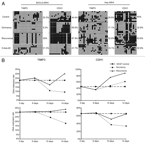
Figure 6. The 5-Aza-dC and /or TSA treatment regulates ovarian cancer cell colony formation and the expression of angiogenesis-related genes. (AandC) Colony number of four groups (recurrence, 5-Aza-dC, TSA, and 5-Aza-dC + TSA) in SKOv3-ARHI (A) and Hey-ARHI (C) cells. *Compared with the recurrence group, P < 0.05. (BandD) Results of 10 genes that showed significant changes after 5-Aza-dC and/or TSA treatment in the recurrence groups measured by real-time RT-PCR were presented for SKOv3-ARHI cells (B) and in Hey-ARHI cells (D). *Compared with the recurrence group, P < 0.05. Independent experiments were repeated at least three times to confirm the reproducibility of the results.
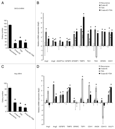
Figure 7. Induction of ARHI induces tumor dormancy in human ovarian cancer xenografts. (A) The control and dormancy groups were provided without DOX or with DOX in their drinking water. In the recurrence group, DOX was withdrawn after 32 d of treatment. Arrowheads indicate the day of DOX withdrawal. The results shown were from three independent experiments. *Compared with the control group, P < 0.01. ##Compared with the recurrence group, P < 0.01. (B) LC3-I, LC3-II and ARHI expression in the three groups were detected by Western Blot. *Compared with the control group, P < 0.05. #Compared with the recurrence group, P < 0.05 (C) PCNA (a cell proliferation marker) and CD31 (an angiogenesis marker) expression detected by immunohistochemistry.
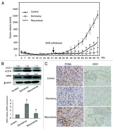
Figure 8. TIMP3 and CDH1 are upregulated in dormancy and downregulated in recurrent SKOv3-ARHI xenografts. (A) The expression of CDH1 and TIMP3 in xenografts of three groups was measured by real-time PCR. *Compared with the control group, P < 0.05. #Compared with the dormancy group, P < 0.05. (BandC): TIMP3 and CDH3 expression in three groups were detected by western blot (B) and by immunohistochemistry (C).
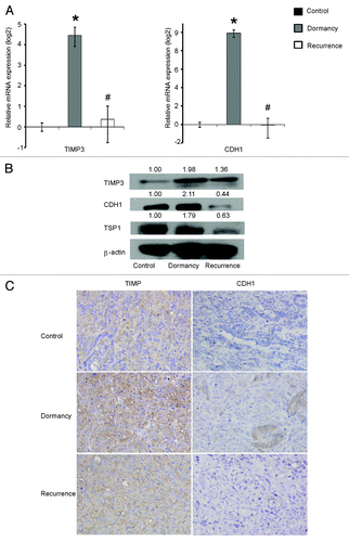
Figure 9.TIMP3 and CDH1 expression is related to DNA methylation in vivo. Methylation status of TIMP3 and CDH1 in the xenografts was measured by bisulfite sequencing. The calculation methods were the same as in .