1,137
Views
49
CrossRef citations to date
0
Altmetric
Research Paper

NANOG promoter methylation and expression correlation during normal and malignant human germ cell development

, , , , &
Pages 114-122 | Received 23 Jul 2010, Accepted 29 Aug 2010, Published online: 01 Jan 2011

Figures & data

Figure 1 Luciferase assay in 2102EP embryonal carcinoma cells. (A) Relative luciferase activity of the NRR (pGL3-NRR), the in vitro methylated NRR (pGL3-NRR M.SssI), the NRR with mutated OCT3/4-SOX2 binding site (mOS) and in vitro methylated NRR with mutated OCT3/4-SOX2 binding site (mOS M.SssI). Untransfected (2102EP u) and “empty” vector transfected cells (pGL3basic) served as controls. (B) NRR methylation level in JKT-1 cells before (untreated) and after treatment with 10 µM 5-aza-2'-deoxycytidine (5-aza). Filled circles indicate methylated CpGs, empty circles indicate demethylated CpGs. (C) RT-PCR analysis of NANOG-expression before (untreated) and after treatment with 5-aza (5-aza).
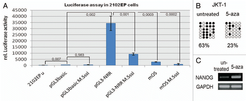
Figure 1 Luciferase assay in 2102EP embryonal carcinoma cells. (A) Relative luciferase activity of the NRR (pGL3-NRR), the in vitro methylated NRR (pGL3-NRR M.SssI), the NRR with mutated OCT3/4-SOX2 binding site (mOS) and in vitro methylated NRR with mutated OCT3/4-SOX2 binding site (mOS M.SssI). Untransfected (2102EP u) and “empty” vector transfected cells (pGL3basic) served as controls. (B) NRR methylation level in JKT-1 cells before (untreated) and after treatment with 10 µM 5-aza-2'-deoxycytidine (5-aza). Filled circles indicate methylated CpGs, empty circles indicate demethylated CpGs. (C) RT-PCR analysis of NANOG-expression before (untreated) and after treatment with 5-aza (5-aza).

Figure 2 (A and B) Immunohistochemical staining of fetal germ cells for c-KIT (A) and MAGE-A4 (B). Arrowheads indicate cells positive for the respective protein. Stained cells were extracted by laser micro-dissection for DNA isolation and sodium-bisulfite sequencing. Scale bar: 500 µm. (C–G) NRR DNA-methylation pattern of germ cells at developmental stages indicated. Adult spermatogonia (E) were isolated by laser micro-dissection. DNA of adult testis tissues (F) was isolated from whole tissue lysates, while sperm DNA was isolated from ejaculates (G). (H) Positive control (M.SssI treated DNA from TCam-2 cells). Filled circles indicate methylated CpGs, empty circles indicate demethylated CpGs.
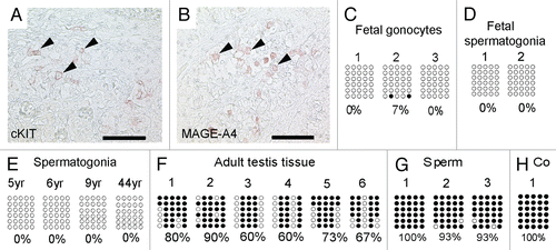
Figure 2 (A and B) Immunohistochemical staining of fetal germ cells for c-KIT (A) and MAGE-A4 (B). Arrowheads indicate cells positive for the respective protein. Stained cells were extracted by laser micro-dissection for DNA isolation and sodium-bisulfite sequencing. Scale bar: 500 µm. (C–G) NRR DNA-methylation pattern of germ cells at developmental stages indicated. Adult spermatogonia (E) were isolated by laser micro-dissection. DNA of adult testis tissues (F) was isolated from whole tissue lysates, while sperm DNA was isolated from ejaculates (G). (H) Positive control (M.SssI treated DNA from TCam-2 cells). Filled circles indicate methylated CpGs, empty circles indicate demethylated CpGs.

Figure 3 Immunohistochemical staining of testis sections at week 19 (A and C) and 24 (B and D) of gestation for OCT3/4 (A and B) and SOX2 (C and D). Arrows indicate cells positive for OCT3/4. Scale bar: 50 µm.
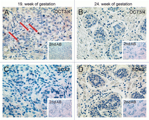
Figure 3 Immunohistochemical staining of testis sections at week 19 (A and C) and 24 (B and D) of gestation for OCT3/4 (A and B) and SOX2 (C and D). Arrows indicate cells positive for OCT3/4. Scale bar: 50 µm.

Figure 4 Immunohistochemical staining detecting global DNA-methylation in different testis samples using 5-mC antibody. (A) Red arrows point at spermatogonia of a testis from 2-year-old patient being positive for 5 mC antibody. (B) Testis section of a 6-year-old patient shows global hypermethylation of spermatogonia, too. (C and D) Spermatids and sperm of donors age 22 and 29 show global hypomethylation (arrowheads). Scale-bars: A and B: 200 µm; C and D: 50 µm.
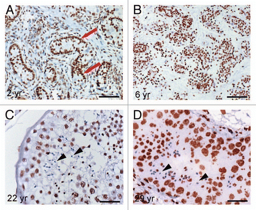
Figure 4 Immunohistochemical staining detecting global DNA-methylation in different testis samples using 5-mC antibody. (A) Red arrows point at spermatogonia of a testis from 2-year-old patient being positive for 5 mC antibody. (B) Testis section of a 6-year-old patient shows global hypermethylation of spermatogonia, too. (C and D) Spermatids and sperm of donors age 22 and 29 show global hypomethylation (arrowheads). Scale-bars: A and B: 200 µm; C and D: 50 µm.

Figure 5 Graphical illustration of the NRR DNA-methylation pattern in indicated GCT samples (A–F). Filled circles indicate methylated CpGs, empty circles indicate demethylated CpGs. (G) Diagram of relative NRR methylation levels of germ cells and GCTs.
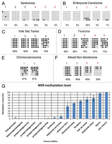
Figure 5 Graphical illustration of the NRR DNA-methylation pattern in indicated GCT samples (A–F). Filled circles indicate methylated CpGs, empty circles indicate demethylated CpGs. (G) Diagram of relative NRR methylation levels of germ cells and GCTs.

Figure 6 (A) NRR-methylation analysis of GCT cell lines. Filled circles indicate methylated CpGs, empty circles indicate demethylated CpGs. (B) qRT-PCR anaylsis of NANOG expression in GCT and control tissues (red colored samples in ) and corresponding cell lines. (C) RT-PCR and (D) western blot of cell lines indicated detecting expression and protein level of NANOG.
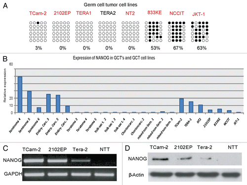
Figure 6 (A) NRR-methylation analysis of GCT cell lines. Filled circles indicate methylated CpGs, empty circles indicate demethylated CpGs. (B) qRT-PCR anaylsis of NANOG expression in GCT and control tissues (red colored samples in Fig. 3A) and corresponding cell lines. (C) RT-PCR and (D) western blot of cell lines indicated detecting expression and protein level of NANOG.
Supplemental material

Additional material

Download Zip (2.2 MB)

Reprints and Corporate Permissions

Please note: Selecting permissions does not provide access to the full text of the article, please see our help page How do I view content?

To request a reprint or corporate permissions for this article, please click on the relevant link below:

Academic Permissions

Please note: Selecting permissions does not provide access to the full text of the article, please see our help page How do I view content?

Obtain permissions instantly via Rightslink by clicking on the button below:

If you are unable to obtain permissions via Rightslink, please complete and submit this Permissions form. For more information, please visit our Permissions help page.