Figures & data
Figure 1 Acta2HpaIIM transgene structure and expression. (A) Schematic map of the Acta2HpaIIM 6.4 kb transgene. Rectangles represent exons or coding sequences. (B) Semi-quantitative RT-PCR analysis of transgene expression in the two independent lines S13 and Z. St, stomach; SI, small intestine; Co, colon; Ao, aorta; VG, vesicular gland; Bl, whole blood cells.
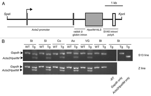
Figure 2 Organomegaly in a subset of Acta2HpaIIM mice. (A) Organ wet weight in WT controls and transgenics of the S13 and Z lines. Data points represent individual mice. Statistical differences are in comparisons with corresponding WT (*p < 0.05; **p < 0.01). Co, colon; SI, small intestine; VG, vesicular gland. (B and C) Examples of hyperplasia of the vesicular gland (arrows) in WT control and transgenic, respectively.
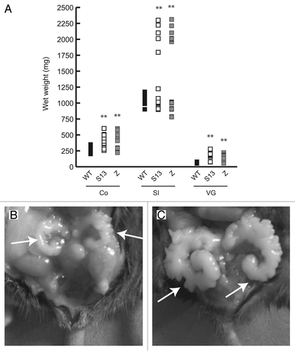
Figure 3 Acta2HpaIIM mice tumors. (A) Representative example of transgene expression in transgenic organs and tumors. VG, vesicular gland; SI, small intestine; N, normal size; H, hyperplastic; Adj, macroscopically normal tissue adjacent to a tumor; IC, intestinal carcinoma; He, hemoangioma. (B–D) Examples of transgenic tumor histology (hematoxylin/eosin). (B) Intestinal carcinoma. An area of well differentiated carcinoma is visible in the lower part, left of arrows (40× magnification). (C) Hemoangioma. The tumor mass surrounds blood vessels with abnormal architecture (arrow; 100×). (D) Lymphoma. Cells with enlarged nuclei (examples indicated by arrows, left) and phagocytes (arrowhead, right) are visible (200×).
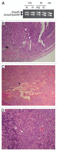
Figure 4 Global DNA hypermethylation in Acta2HpaIIM mice tumors. (A) Total 5mdC normalized as percent of dG in organs and tumors (average ± SD). Organs: white bars, WT controls; grey bars, transgenic normal size organs; black bars, transgenic hyperplastic organs (n = 5 in each group). Co, colon; SI, small intestine; VG, vesicular gland. Tumors: white and black bars, adjacent tissue and tumor mass, respectively. He, hemoangioma (n = 5); IC, intestinal carcinoma (n = 3); KA, kidney adenoma (n = 3); Ly, lymphoma (n = 5). Statistical significance: *p < 0.05; **p < 0.01. (B and C) quantitative methylation-sensitive PCR analysis of LINE-1 methylation status. HpaII endonuclease is sensitive to methylation of the underlined (internal and external) cytosine residues in CCGG. MspI is an isoschizomer of HpaII but is sensitive to methylation of the external cytosine residue (CCGG). Amplification indicates methylation at the corresponding endonuclease site residue(s). (D and E) as in (B and C) but for MusD elements. Adj., apparently normal tissue adjacent to tumor mass; SM, skeletal muscle; for other symbols see (A).
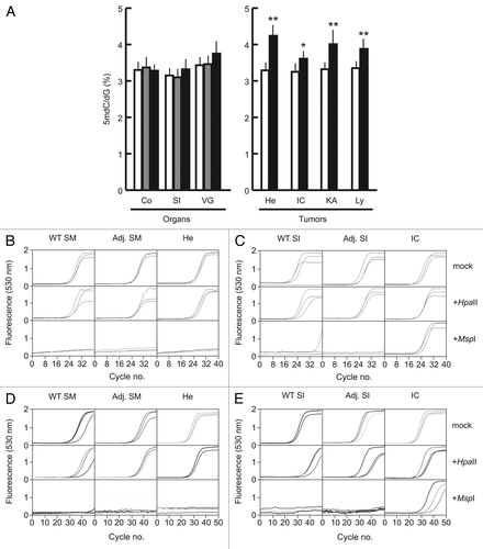
Figure 5 DNA methylation status of tumor suppressor genes and intragenic CpG islands in Acta2HpaIIM mice tumors. (A) Promoter bisulfite DNA methylation analysis and quantitative RT-PCR expression of selected genes in a hemoangioma and intestinal carcinoma. Gene symbols are on the left. In DNA methylation graphs, numbers on the vertical axis indicate the analyzed sequence position relative to the TSS (bp). Ticks represent CpG dinucleotides, arrows and arrowheads indicate HpaII site CpG (CCGG) and external cytosines of HpaII sites (CCGG), respectively. Horizontal bars represent the extent of methylation of the corresponding residues in the clones analyzed. In expression graphs, values are relative to Gapdh RNA levels (average ± SD). White bars, WT controls; grey bars, transgenic adjacent tissue; black bars, transgenic tumor mass. Adj., apparently normal transgenic tissue adjacent to tumors; He, hemoangioma; IC, intestinal carcinoma; SI, small intestine; SM, skeletal muscle. Statistical significance: *p < 0.05; **p < 0.01. (B) methylation status of a sequence located 3′ to Cdkn2a. Numbers on the vertical axis indicate the distance from the 3′ end of Cdkn2a. (C) methylation status of a negative control human MT2A CpG island. For graph symbols in (B and C) see (A).
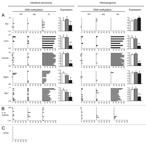
Figure 6 Promoter DNA methylation status of non-tumor suppressor genes in Acta2HpaIIM mice tumors. Symbols same as in legend of .
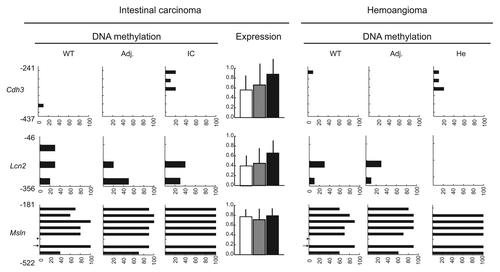
Table 1 Tumor type and frequency in transgenics from S13 and Z lines, and WT controls (n = 40 per group)