Figures & data
Figure 1 Representative photomicrographs of matched Hematoxylin and Eosin stained frozen sections of benign (A) and malignant (B) prostatic tissues and CD31 immunostained frozen sections of benign (C, E and F) and malignant (D, H–J) prostatic tissues prepared for LMD. The benign prostatic tissue shows stromal and glandular hyperplasia and the malignant prostatic tissue shows well-differentiated adenocarcinoma. Endothelial cells are highlighted by CD31 immunostaining of matched benign (C) and malignant (D) prostate frozen tissue on a PEN membrane slides. It was a common observation that malignant prostatic tissues had higher microvascular density than benign prostatic tissues. (E and H) represent CD31 immunostained tissue sections before microdissection. (F and I) represent outlined endothelium to be dissected. (G) represents captured endothelium from the slide in a collection cap. (J) represents the remaining tissue sections after microdissection.
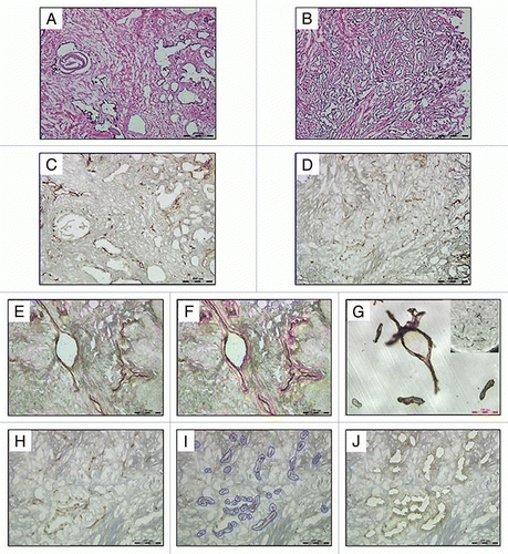
Figure 2 CYP24A1 promoter DNA methylation of endothelium associated with benign and malignant prostate tissues. (A) Diagram of the CYP24A1 promoter CpG-rich region. Twenty individual clones were sequenced from the CYP24A1 promoter region and shown from a representative patient sample (left). Positive (+) and negative (−) methylation controls from PC-3 and DU145, respectively, are shown (right). The methylation percentage indicates total proportion of methylated CpGs in the indicated CYP24A1 promoter region for each sample, taking into account all sequenced alleles. (B) Results indicate that the CYP24A1 promoter is methylated in the CD31 enriched endothelium associated with human prostate tumor compared to benign prostate (p = 0.045, paired, two-tailed Student's t-Test). Mean ± SEM values are shown. Matched patient pair of prostate tumor and benign prostate endothelium are shown with symbols: Patient #1 (■), Patient #2 (△), Patient #3 (○), Patient #4 (◊), Patient #5 (●) and Patient #6 (▲). (C) Lack of correlation of CYP24A1 promoter region 2 methylation between CD31+ endothelium from LMD and remaining epithelium/stroma from the de-vascularized sections.
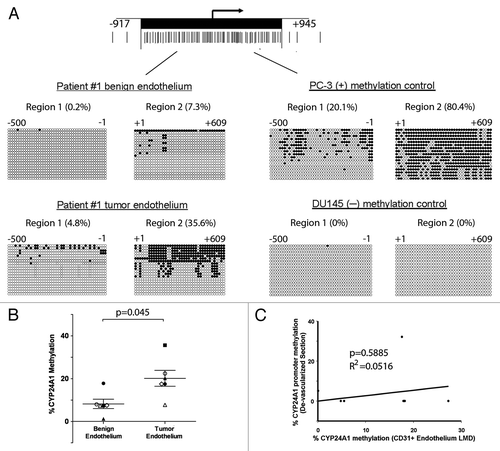
Table 1 Laser microdissection of CD31+ cells from prostate tumor and matched histologically benign prostatic tissues from radical prostectectomy
Table 2 Percent CYP24A1 promoter methylation of CD31+ endothelium from matched benign and tumor prostatic tissues