Figures & data
Table 1. Genes that control salivary gland migration and/or lumen size
Figure 1. Rho1 is required for salivary gland migration and correct lumen size. In Rho11B heterozygous embryos (A and B), the salivary gland lumen elongates and narrows at the proximal end as the gland migrates (A and B, arrows). In Rho11B homozygous embryos, the salivary gland fails to migrate and elongate, and lumen width is expanded throughout the gland (C, arrow). Embryos were stained for dCREB-A (green) to label salivary gland nuclei, E-cadherin (E-cad) (white) to label the lumen and β-galactosidase (not shown) to distinguish heterozygous from homozygous embryos. The lumen in all panels is outlined in red and shaded in red. Scale bar in A represents 5 μm.
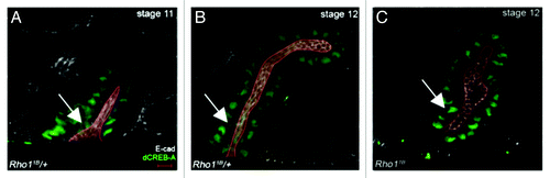
Figure 2. Salivary gland cells rearrange and transition from a pseudostratified to a simple epithelium during collective migration. In wild-type embryos (A-C), salivary gland cells rearrange as indicated by a decrease in the number of nuclei that surrounds the lumen. In Rho11B homozygous embryos at stage 12 (D), salivary gland cells fail to rearrange. In salivary glands of stage 11 wild-type embryos (E), the epithelium is pseudostratified with nuclei located at different positions along the length of the cell. By stage 12 in wild-type embryos (F), the salivary gland epithelium has transitioned to a simple epithelium with nuclei located at a similar position in all cells. All embryos shown were stained for dCREB-A (green in A-D; red in E and F) to label salivary gland nuclei; embryos in A-D were also stained for E-cadherin (E-cad; white) and those in E and F for aPKC (green) to label the lumen and Neurotactin (Nrt; white) to label the basolateral membrane. Scale bar in A represents 3 μm.
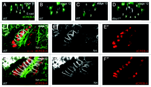
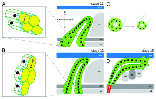
Figure 3. Cellular processes that control salivary gland migration and lumen size. At stage 11, all salivary gland cells have invaginated and the gland is oriented dorsally. Cells in the proximal part of the gland are columnar shaped with isotropic apical domains, and form a pseudostratified epithelium with nuclei at different positions along the apical-basolateral axis of the cell (A). As the salivary gland migrates dorsally at stage 12, the distal tip contacts the overlying circular visceral mesoderm (CVM) and proximal gland cells begin to change shape from columnar to cuboidal (B). It is also during stage 12 that proximal gland cells transition from a pseudostratified to a simple epithelium, apical domains elongate in the direction of migration (B) and cells rearrange to form a narrower tube (C). By stage 13, distal gland cells extend basal membrane protrusions as they migrate along the overlying CVM and underlying fat body (FB) and somatic mesoderm (SM) (D). Salivary gland cells are depicted in green and salivary duct cells in red. D: dorsal; V: ventral; A: anterior; P: posterior. Di: distal; Pr: proximal. VE: ventral ectoderm; VNC: ventral nerve cord; NB: neuroblasts. Diagram is not drawn to scale.