Figures & data
Table 1. Optimized conditions for Drosophila LV-SEM using the FEI Quanta 450
Figure 1. Effects of spot size, working distance, and pressure on LV-SEM images. Bar = 10 µm for each set of images. The contrast was adjusted post-collection in this and the remaining figures, except where noted (see Materials and Methods). (A–E) Effect of spot size on the w1118 eye; the same region is shown in each panel; shot at 1000× magnification. Spot sizes were 2.0 (A); 2.5 (B); 3.0 (C); 3.5 (D); and 4.5 (E) as indicated. Resolution is slightly better at lower spot sizes, but the signal is lower and noise more apparent. Spot = 3.5 balances good resolution with low noise and sample damage. (F-H) Thoracic bristles of w1118 were scanned at 2000× and 15 kV, with spot sizes 2.5 (F), 3.5 (G), and 5.0 (H) as indicated. The same region is shown in each panel. Using spot = 5.0 leads to singed hairs (arrowhead) and charging on the bristle sockets, problems not observed with spot = 3.5. (I–J) Increased working distance (WD) provides increased depth of field. Focus is set on the upper surface of a w1118 haltere in both panels, taken at 500×. Note the underlying thorax has better focus in (J) (WD = 14.8 mm) than in (I) (WD = 5.0 mm). (K–M) Variation in chamber pressure, using orbital bristles taken at 2500× and shown with original contrast. The same bristle socket is shown at 0.30 (K), 0.53 (L), and 0.68 Torr (M).
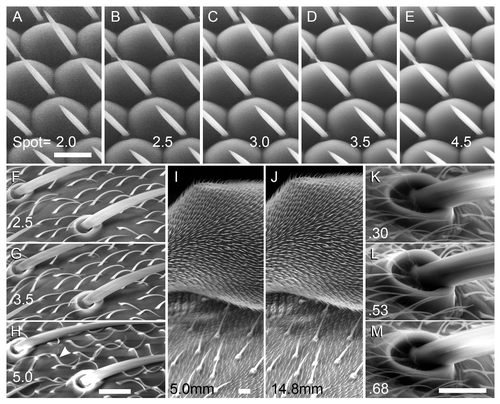
Figure 2. Effect of accelerating voltage on the w1118 eye and the sex comb of the male foreleg. All panels were shot at 1000×; bar = 10 µm for all. A-D, w1118 eye; the same region is shown in each panel. Voltages were 10 (A); 15 (B); 20 (C); and 25 kV (D). Appearance of ommatidia changes substantially as voltage varies; the 15 kV image most resembles a conventional high-vacuum SEM image. (E–F) the striations of the sex comb bristles are clearly seen at 7.5 kV (E), but not at 15 kV (F).
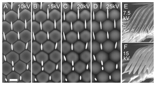
Figure 3. Examples of wild type structures at varying magnifications in w1118 flies. (B, D and E) were scanned in large format (2048 pixel width). Beam = 15 kV for (A, B, E and F). (A) Whole female mounted in CCC; bar = 1 mm; shot at 20×. (B) Male genitalia (ventral); bar = 50 µm; shot at 250×. (C) Male wing margin (distal is up); bar = 10 µm; shot at 500× and 7.5 kV. (D) Male tarsal claw and pulvillus; bar = 10 µm; shot at 2000× and 10 kV. (E) Sensory hair bed on the ventral anterior side of the thorax near the head (male; anterior is up). Bar = 10 µm; shot at 1500×. (F) Proboscis; bar = 50 µm; shot at 340× and spot = 3.0.
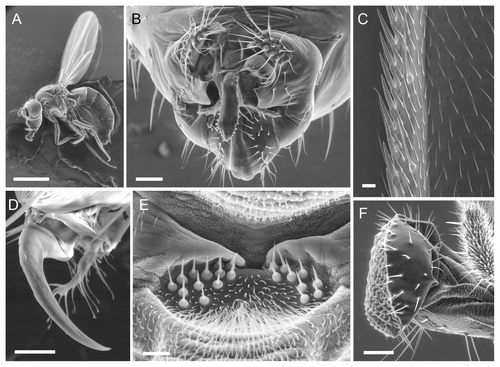
Figure 4. Analysis of mutant phenotypes using LV-SEM. Bars = 10 µm for each pair of images. (A–B) orbital bristles from dorsal head of w1118 (A) and sn (B) males shot at 750×, 10 kV. (C–D) retina of w1118 (C) and GMR-GAL4 > Mi[Hto-WP]QYE (D), shot at 1000× and 2048 pixel width. (E–F) Equivalent regions of w1118 (E) and ms1096w-GAL4 > Mi[Hto-WP]VRC male wings (F), shot at 1500×, 7.5 kV. Note multipronged wing hairs in (F). (G–H) Wing surface with L3 vein and campaniform sense organ (center), from w1118 (G) and ms1096w-GAL4 > Mi[Hto-WP]NAW (H), shot at 1000×, 7.5 kV. In (H), note the hairs are eliminated from this region of the wing, and the cuticle is abnormal.
![Figure 4. Analysis of mutant phenotypes using LV-SEM. Bars = 10 µm for each pair of images. (A–B) orbital bristles from dorsal head of w1118 (A) and sn (B) males shot at 750×, 10 kV. (C–D) retina of w1118 (C) and GMR-GAL4 > Mi[Hto-WP]QYE (D), shot at 1000× and 2048 pixel width. (E–F) Equivalent regions of w1118 (E) and ms1096w-GAL4 > Mi[Hto-WP]VRC male wings (F), shot at 1500×, 7.5 kV. Note multipronged wing hairs in (F). (G–H) Wing surface with L3 vein and campaniform sense organ (center), from w1118 (G) and ms1096w-GAL4 > Mi[Hto-WP]NAW (H), shot at 1000×, 7.5 kV. In (H), note the hairs are eliminated from this region of the wing, and the cuticle is abnormal.](/cms/asset/bf63d76c-2ca5-4137-9062-ce0d349f4672/kfly_a_10920525_f0004.gif)