Figures & data
Figure 1. The green fluorescent color detected in (A) non-infiltrated petal and (B) the pollen grain in non-infiltrated under the fluorescent UV microscope.
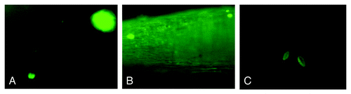
Figure 2. GUS expression in agroinfiltrated gerbera flower in (A) dark pink and (B) white flower colors.
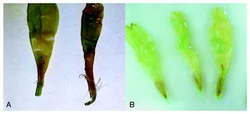
Table 1. Level of blue color intensities for GUS transient expression in agroinfiltrated gerbera flower petals
Figure 3. Cross sections in gerbera petals under 40× light microscope for Nonagroinfiltrated flower (A) with uniform color, agroinfiltrated petal with dfr gene (B), f3′5'h/dfr(C) and f3′5'h(D) showing ranging of color variation petal compared with the non-infiltrated.
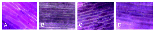
Figure 4. Arrows pointed to the blue stigma (A) blue pollen grain (B) after agroinfiltration with color involved genes
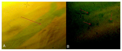
Figure 5. The color petal's bases of the (A) non-agroinfiltrated gerbera flower controlled compared with gerbera with (B)dfr gene, (C) a combination of dfr and f3′5'h gens showing dark bases color and (D)f3′5'h gene
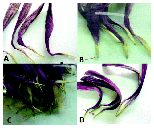
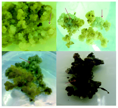
Figure 6. Light and dark blue gerbera callus generated after the transformation with the f3'5'h gene.