Figures & data
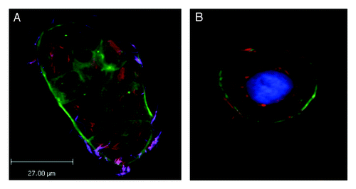
Figure 1. Immunofluorescence micrographs showing images of Caco-2 cells infected with F. nucleatum strain EAVG_002 for 24 h, showing localization of bacteria in the perinuclear region. Strain EAVG_002 is an example of a highly invasive strain isolated from an inflamed intestinal biopsy taken from an IBD patient and has been extensively characterized.Citation3,Citation8,Citation47 (A) This merged image shows differentially stained bacteria (see ref. Citation8 for differential staining method used) allowing clear delineation between bacteria that have invaded and are internalized within the imaged group of host cells and bacteria that remain outside of these host cells; actin is labeled with Phalloidin 488 (green), internalized bacteria are orange (Cy3) and bacteria external to the cells appear purple (Cy3 + Alexa 350). (B) Merged image showing detail of a representative host cell from sample taken from same experimental set as for (A) except that for labeling purposes this time differential staining was not done and instead samples were stained with DAPI (blue) and Phalloidin 488 (green) to reveal the host cell nuclei and actin cytoskeleton respectively and all bacteria were stained orange using Cy3.