2,675
Views
33
CrossRef citations to date
0
Altmetric
Review

Bacterial symbioses of the medicinal leech Hirudo verbana

&
Pages 322-331 | Published online: 10 May 2012

Figures & data

Figure 1. Major alimentary structures and microbial interactions of Hirudo verbana. Drawing demonstrating the anatomy of Hirudo verbana showing the major structures of the alimentary system including the crop, lateral crop ceca, nephridia and bladders, intestinum, and anus. Inset images illustrate microbial interactions within the bladder (A), along the crop epithelium (B) and through the crop at large (C) and are as follows: A) FISH image showing the distribution of bacteria within the bladder lumen (colors correspond to probes targeted as follows: green = β-Proteobacteria, red = Bacteroidetes, blue = Bacteria); (B) FISH image of the Rikenella-like bacterium (red) along the crop epithelium, epithelial cell nuclei counter-stained with DAPI; (C1) composite image of Aeromonas (red) associated with the cell surface of a leech hemocyte, nucleus counter-stained with DAPI; (C2) FISH image of mixed microcolonies showing both Aeromonas (green) and the Rikenella-like bacterium (red), erythrocytes from a blood meal auto-fluoresce green and have darkened interiors; (C3) fluorescent image of lectin staining using succinylated wheat germ agglutinin (WGA-S, red) and DAPI counter-staining of bacterial colonies. For images A and B, the following indicators are used: E indicates the tissue epithelium of the bladder (A) or crop (B) and L indicates the lumen.
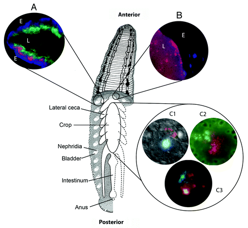
Figure 1. Major alimentary structures and microbial interactions of Hirudo verbana. Drawing demonstrating the anatomy of Hirudo verbana showing the major structures of the alimentary system including the crop, lateral crop ceca, nephridia and bladders, intestinum, and anus. Inset images illustrate microbial interactions within the bladder (A), along the crop epithelium (B) and through the crop at large (C) and are as follows: A) FISH image showing the distribution of bacteria within the bladder lumen (colors correspond to probes targeted as follows: green = β-Proteobacteria, red = Bacteroidetes, blue = Bacteria); (B) FISH image of the Rikenella-like bacterium (red) along the crop epithelium, epithelial cell nuclei counter-stained with DAPI; (C1) composite image of Aeromonas (red) associated with the cell surface of a leech hemocyte, nucleus counter-stained with DAPI; (C2) FISH image of mixed microcolonies showing both Aeromonas (green) and the Rikenella-like bacterium (red), erythrocytes from a blood meal auto-fluoresce green and have darkened interiors; (C3) fluorescent image of lectin staining using succinylated wheat germ agglutinin (WGA-S, red) and DAPI counter-staining of bacterial colonies. For images A and B, the following indicators are used: E indicates the tissue epithelium of the bladder (A) or crop (B) and L indicates the lumen.

Table 1. List of Aeromonas veronii mutants

Reprints and Corporate Permissions

Please note: Selecting permissions does not provide access to the full text of the article, please see our help page How do I view content?

To request a reprint or corporate permissions for this article, please click on the relevant link below:

Academic Permissions

Please note: Selecting permissions does not provide access to the full text of the article, please see our help page How do I view content?

Obtain permissions instantly via Rightslink by clicking on the button below:

If you are unable to obtain permissions via Rightslink, please complete and submit this Permissions form. For more information, please visit our Permissions help page.