1,029
Views
12
CrossRef citations to date
0
Altmetric
Research Paper

Advances in vaccines against neglected tropical diseases

Enhancing physical stability of a recombinant hookworm vaccine through biophysical and formulation studies

, , , , , , & show all
Pages 765-776 | Published online: 12 Apr 2012

Figures & data

Figure 1. (A) SDS-PAGE under reducing conditions containing 2 µg of Na-GST-1 in 50 mM sodium acetate pH 6.0 buffer. Lane A represents reference protein stored at < -50°C and Lanes B-E are replicate samples of the recombinant protein stored at 2–8°C. (B) SDS-PAGE under non-reducing conditions of Na-GST-1 samples. Lanes A and B correspond to supernatant from two protein samples where particulates were observed after freeze thaw. Lanes C and D represent protein samples from lanes A and B after they were centrifuged to isolate particulates and resuspended in sample buffer. (C-D) SE-HPLC of two recombinant Na-GST-1 protein samples. Panel C represents recombinant protein stored at freezing temperature (below -50°C). Panel D represents recombinant protein stored at 2–8°C for 3 mo with a decrease in main peak area, purity and detection of lower molecular weight breakdown products.
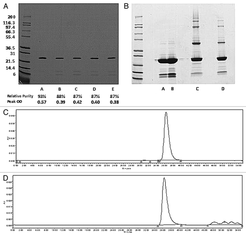
Figure 1. (A) SDS-PAGE under reducing conditions containing 2 µg of Na-GST-1 in 50 mM sodium acetate pH 6.0 buffer. Lane A represents reference protein stored at < -50°C and Lanes B-E are replicate samples of the recombinant protein stored at 2–8°C. (B) SDS-PAGE under non-reducing conditions of Na-GST-1 samples. Lanes A and B correspond to supernatant from two protein samples where particulates were observed after freeze thaw. Lanes C and D represent protein samples from lanes A and B after they were centrifuged to isolate particulates and resuspended in sample buffer. (C-D) SE-HPLC of two recombinant Na-GST-1 protein samples. Panel C represents recombinant protein stored at freezing temperature (below -50°C). Panel D represents recombinant protein stored at 2–8°C for 3 mo with a decrease in main peak area, purity and detection of lower molecular weight breakdown products.

Figure 2. Ezymatic activity of 40 µg Na-GST-1 protein measured by fluorescence assay and reported in relative fluorescence units (RFU) under different treatment conditions: Control = untreated (frozen protein and thawed for use); heat treated at two temperatures (55°C and 100°C) and under reducing conditions with DTT). Results shown are average of duplicate measurements.
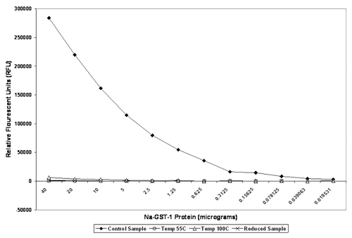
Figure 2. Ezymatic activity of 40 µg Na-GST-1 protein measured by fluorescence assay and reported in relative fluorescence units (RFU) under different treatment conditions: Control = untreated (frozen protein and thawed for use); heat treated at two temperatures (55°C and 100°C) and under reducing conditions with DTT). Results shown are average of duplicate measurements.

Figure 3. (A) The effect of ph on the secondary structure of Na-GST-1 at 10◦C. Each data point represents individual measurements of CD spectra from a single representative sample. (B) Ellipticity at 222 nm as a function of temperature was measured over the pH range of 3–8. Duplicate measurements were taken and the average value displayed. (C) Tryptophan emission fluorescence peak position of Na-GST-1 as a function of pH and temperature. Na-GST-1 at pH 3–8 was heated from 10 – 87.5°C, and the fluorescence emission maximum was determined after excitation at 295 nm. (D) Tryptophan emission fluorescence intensity of Na-GST-1 as a function of pH and temperature. Na-GST-1 at pH 3–8 was heated from 10 – 87.5°C. Error bars are from duplicate measurements.
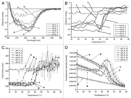
Figure 3. (A) The effect of ph on the secondary structure of Na-GST-1 at 10◦C. Each data point represents individual measurements of CD spectra from a single representative sample. (B) Ellipticity at 222 nm as a function of temperature was measured over the pH range of 3–8. Duplicate measurements were taken and the average value displayed. (C) Tryptophan emission fluorescence peak position of Na-GST-1 as a function of pH and temperature. Na-GST-1 at pH 3–8 was heated from 10 – 87.5°C, and the fluorescence emission maximum was determined after excitation at 295 nm. (D) Tryptophan emission fluorescence intensity of Na-GST-1 as a function of pH and temperature. Na-GST-1 at pH 3–8 was heated from 10 – 87.5°C. Error bars are from duplicate measurements.

Figure 4. (A) Static light scattering as a function of pH and temperature. Na-GST-1 at pH 3–8 was heated from 10 – 87.5°C and the scattering intensity was monitored at 295 nm. Error bars are from duplicate measurements. (B) Binding of ANS to Na-GST-1 as a function of pH and temperature. A 20:1 ANS:protein molar ratio was excited at 372 nm and the fluorescence intensity at 484 was monitored as a function of temperature. Error bars are from duplicate measurements.
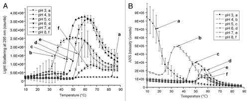
Figure 4. (A) Static light scattering as a function of pH and temperature. Na-GST-1 at pH 3–8 was heated from 10 – 87.5°C and the scattering intensity was monitored at 295 nm. Error bars are from duplicate measurements. (B) Binding of ANS to Na-GST-1 as a function of pH and temperature. A 20:1 ANS:protein molar ratio was excited at 372 nm and the fluorescence intensity at 484 was monitored as a function of temperature. Error bars are from duplicate measurements.

Figure 5. Empirical phase diagram (EPD) derived from biophysical characterization of Na-GST-1. The EPD is prepared from temperature-dependent intrinsic tryptophan fluorescence peak position, peak intensity, extrinsic ANS fluorescent peak intensity, circular dichroism at 222 nm, and static light scattering (see text).
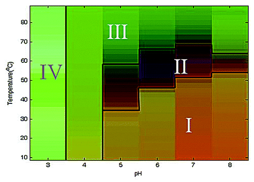
Figure 5. Empirical phase diagram (EPD) derived from biophysical characterization of Na-GST-1. The EPD is prepared from temperature-dependent intrinsic tryptophan fluorescence peak position, peak intensity, extrinsic ANS fluorescent peak intensity, circular dichroism at 222 nm, and static light scattering (see text).

Figure 6. Excipient screening of Na-GST-1 using OD350 measurements. The change in optical density of Na-GST-1 is displayed as a function of time. Selected results are shown as an example. Experiments were conducted at pH 5 and 50°C.
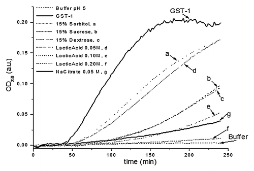
Figure 6. Excipient screening of Na-GST-1 using OD350 measurements. The change in optical density of Na-GST-1 is displayed as a function of time. Selected results are shown as an example. Experiments were conducted at pH 5 and 50°C.

Table 1. Inhibition of aggregation of Na-GST-1 in the presence of various excipients

Table 2. Representative stability testing of Na-GST-1 recombinant protein manufactured in 10% D-glucose, 10 mM Imidazole pH 7.4 buffer and stored at 2–8°C up to 18 mo

Supplemental material

Additional material

Download Zip (190.5 KB)

Reprints and Corporate Permissions

Please note: Selecting permissions does not provide access to the full text of the article, please see our help page How do I view content?

To request a reprint or corporate permissions for this article, please click on the relevant link below:

Academic Permissions

Please note: Selecting permissions does not provide access to the full text of the article, please see our help page How do I view content?

Obtain permissions instantly via Rightslink by clicking on the button below:

If you are unable to obtain permissions via Rightslink, please complete and submit this Permissions form. For more information, please visit our Permissions help page.