Figures & data
Figure 1. The CD11c+ cell purification with MACS. (A) We can see easily that the BMDCs have been purified significantly and approached 95%. (B) BMDCs proliferation under a range of RG doses and BMDCs proliferation under a range of RG+MENK doses.
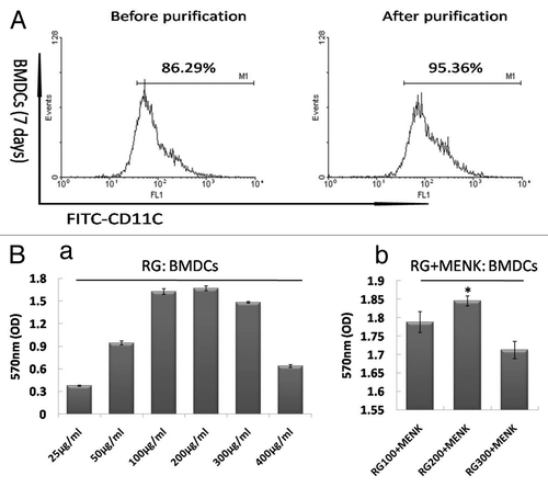
Figure 2. Up-regulation of key surface molecules on BMDCs after treatment with RG and/or MENK for 48h. (A) The cells were respectively collected and stained with mAbs to CD40, CD83, and CD86. Expression of surface markers was analyzed by FCM and was displayed respectively by the single parameter diagram. The values shown in the profiles were the gated %. Results represent the mean ±SD of three independent samples. The images of BMDCs before and after treatment with RG and or MENK (B) by SEM (B) (×3500).
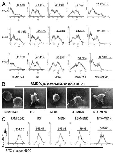
Figure 3. The production of IL-12p40 (a), IL-12p70 (b) and TNF-α (c) by BMDCs after treatment with RG and/or MENK for 48hr by ELISA. After treatment with RG and/or MENK the supernatant from cell cultures was collected and the secreted cytokines were quantified by ELISA. The histograms above showed the levels of IL-12p40, IL-12p70 and TNF-α production .Results represented the mean±SD of three independent experiments samples.
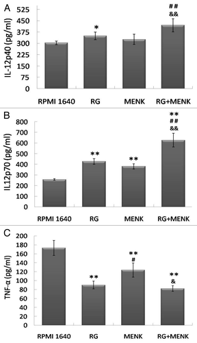
Figure 4. The CD3+ CD8+ T cell purification with MACS. The CD8+T cells from the splenocytes were separated and purified with magnetic beads under sterile condition t. The purity of CD11c+ cells were examined and the percentage were approached 10% to 12%. Followed by purification with MACS, the CD3+ CD8+ T cells were enriched and the results of FCM showed that the purity approached 85%.
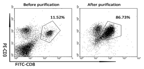
Figure 5. The co-cultured BMDCs and CD8+ T cells (A) in a range of different ratio under a light microscope (200 ×). The purified CD8+T cells were incubated with BMDCs (BMDCs/CD8+ T cells in a ratio of 1:5, 1:10, 1:20 and 1:40) for 5d (B). The BMDCs/CD8+T cells at ratio 1:10 showed the best effect on driving CD8+ T cells proliferation. Up-regulation of CD28, FasL and Prf on CD8+ T cells after treatment with RG and/or MENK BMDCs for 5d (C). The expression of markers was analyzed by FCM and was displayed respectively by the single parameter diagram. The values shown in the profiles were the gated %. Results represent the mean ±SD of three independent samples.
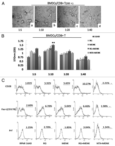
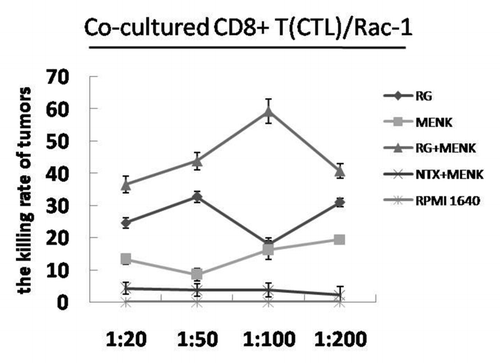
Figure 6. CTL cytotoxicity. The CTL cells were incubated with Rac-1cells (in a ratio of 1:20, 1:50, 1:100 and 1: 200) for 5d. At the ratio 1:100, The CTL cells treated with RG + MENK showed the best cytotoxicity to kill tumor cells( p <0.01).