Figures & data
Figure 1. After cultured with GM-CSF and IL-4 for 6 d, the purity of CD11c+ cells were examined by FACS and the percentage were over 86%. Then purified by MACS, the CD11c+ cells were enriched and the result of FACS approached 95%. FSC/SSC plot was shown in order to get an impression on the purity of the isolated cells.
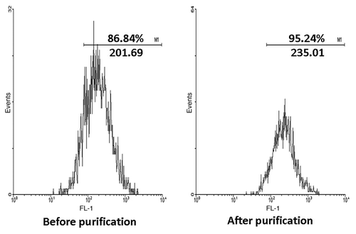
Figure 2. Determination of BMDCs proliferation at different time point (A) and at different concentration of NGP (B) by MTS method.
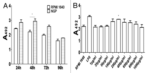
Figure 3. The morphology(×200) under light microscope showed the change of BMDCs. (A) RPMI 1640, (B) NGP.
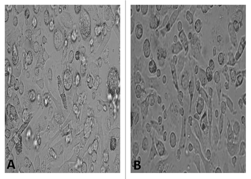
Figure 4. Scanning electron microscopy showed BMDCs morphology. Most of the BMDCs treated with RPMI 1640 were small and round with smooth rim shown in (×5000). However, BMDCs treated with NGP have rough cell surface with many folds and different types of protrusions shown in (×2000).
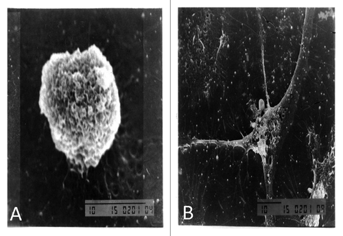
Figure 5. Transmission electron microscopy revealed BMDCs morphology, surface, cytoplasm and organelles. After cultured with NGP for 48 h, the majority of cells had a more irregular surface with more cytoplasmic projections, less vacuoles (heavy arrow) and lysosomes (thin arrow). (A) RPMI1640, (B) NGP (×5,000).
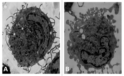
Figure 6. NGP decreased ACP activity inside the BMDCs. All data of ACP activity were shown as means ± S.E.M. (n = 3).
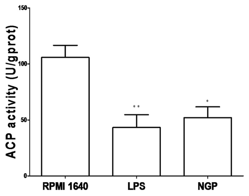
Figure 7. Cellular immunohistochemistry of phagocytosis by BMDC. The BMDCs in different groups were stained with DAB kit. The BMMCs were observed with an inverted phase contrast microscope (CMS GmbH Light Microscopes, Leica Microsystems) (×400).The immature BMDC filled with DAB precipitate could be seen clearly.
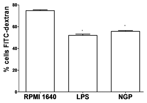
Figure 8. Comparison of FITC-dextran uptaking by immature and mature BMDCs. The BMDCs in different groups were prepared and compared in their ability to uptake FITC-dextran. A representative of three independent experiments was shown. The cells with lower FITC-dextran uptake were preincubated with LPS or NGP. For the analysis of dextran uptaking, the data for controls incubated at 4°C should be subtracted from the 37°C data. All data are represented as means ± S.E.M.(n = 3). *p < 0.05 vs. these in RPMI 1640. **p < 0.01 vs. those in RPMI 1640.
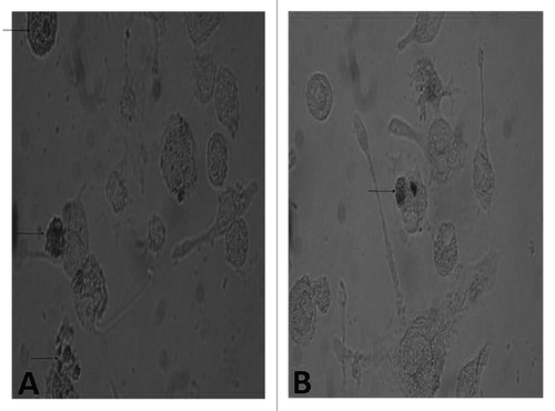
Figure 9. (A) NGP enhanced the phenotypic maturation of BMDC. The BMDC were phenotypically assessed using FACS analyses to determine the expression levels of specific cell surface molecules, as shown in the percentage of expression. NGP clearly increased the expression of CD40, CD80, CD83, CD86 and MHC-II (a representative of three independent experiments). (B) NGP enhanced the phenotypic maturation of murine DCs. All data were shown as means ± S.E.M.(n = 3). *p < 0.05 vs. these in RPMI 1640. **p < 0.01 vs. those in RPMI 1640.
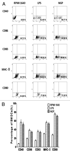
Figure 10. The BMDCs matured in the presence of LPS or NGP produce more IL-12p70 and TNF-α. Cytokines IL-12p70 and TNF-α secretion were tested by ELISA from cultured supernatants of 5 × 10⁴ cells/well of immature and mature BMDC. Results were expressed as the mean ± SEM of triplicate samples. NGP led to an approximate twice of production in IL-12p70 with *p < 0.05 vs. these in RPMI 1640 and TNF-α with **p < 0.01 vs.those in RPMI 1640.
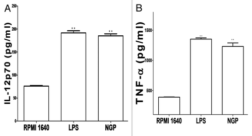
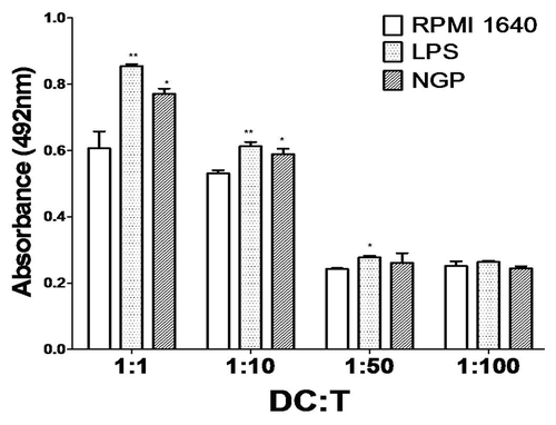
Figure 11. Mixed lymphocyte reaction assay. Dendritic cells derived from bone marrow were co-cultured with the lymphocytes for 3 d at the ratios of 1:1, 1:10, 1:50, 1:100 and proliferation of lymphocytes was measured by MTS assay. All data were shown as means ± S.E.M. (n = 3). *p < 0.05 vs. these in RPMI 1640. **p < 0.01 vs. those in RPMI 1640.