Figures & data
Figure 1. Findings of an early-phase contrast-enhanced CT (CT) scan. (A) Contrast-enhanced CT scan before vaccination shows a 68 × 51-mm tumor with multiple intrahepatic tumors (arrow) and a 44 × 30-mm tumor invading the right atrium (arrowhead). (B) Contrast-enhanced CT after the second vaccination showing multiple low-density areas in the liver, indicating extensive tumor necrosis (arrow). By contrast, a tumor thrombus in the right atrium increased to a 83 × 50-mm tumor (arrowhead). (C) Clinical course from the beginning of GPC3 peptide vaccination. Approximately 1 week after the first vaccination, the patient began reporting general fatigue and showed intermittent fever. Inflammatory and hepatic parameters were elevated (CRP: pink line, AST: red line, T-bil: green line). The abnormal laboratory parameters improved after observation. On day 9 after the second vaccination, the patient was admitted to our hospital as an emergency due to fever and general fatigue, which were similar to his previous symptoms. One day after hospitalization, the inflammatory and hepatic parameters were remarkable. Inflammatory and hepatic parameters improved 1 week after hospitalization. However, his status gradually worsened, and he died on day 30 after the second vaccination. (D) Immunological monitoring of the GPC3 peptide-specific T cell responses. Ex vivo IFN-γ enzyme-linked immunospot (ELISPOT) assays against GPC3 in 5 × 10⁵ peripheral blood mononuclear cells (PBMCs) were performed before and after vaccination. The ∆ spot number indicates the number of GPC3 peptide-specific cytotoxic T-lymphocytes (CTLs). The number of interferon (IFN)-γ positive spots increased from 0 to 84 after the second vaccination.
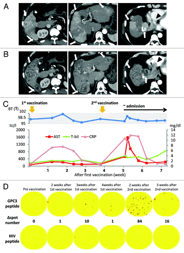
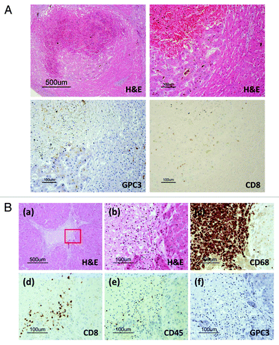
Figure 2. (A) Pathological findings of liver biopsy specimens before vaccination. A microscopy image of a hematoxylin and eosin (H&E)-stained section shows well-differentiated hepatocellular carcinoma (HCC). Immunohistochemical staining for GPC3 and HLA class I showed positivity in the cytoplasm and membranes of carcinoma cells, respectively. No CD8-positive T cells were observed in carcinoma tissue before vaccination. (B) Macroscopic findings of the liver and heart before formalin fixation at the time of autopsy. Most liver tumors had a necrotic area (arrow). A tumor thrombus occupied most of the right atrium (arrow). (C) Pathological findings of the autopsy specimen. (a) Microscopic images of H&E-stained sections showing central necrosis of carcinoma tissue, whereas a cirrhotic nodule adjacent to the carcinoma tissue was not necrotic. (b) Magnified image of the area enclosed within the white box in (a) showing that cancer cells exhibited a morphology (left) different from that of cirrhotic cells (right). (c) CD8-positive T cells (brown) infiltrated the carcinoma cells accompanied by necrosis. In contrast, no infiltration of CD8-positive T cells was detected within the cirrhotic nodule. (d) Magnified image of the area enclosed within the red box in (a) showing necrosis and viable carcinoma cells. (e) Positive immunohistochemical GPC3 staining was observed in only the cytoplasm of carcinoma cells. (f) CD8-positive T cells infiltrated the necrotic area and carcinoma tissue.
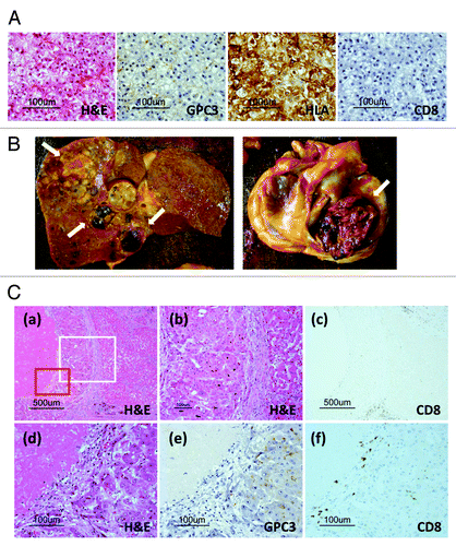
Figure 3. Pathological findings in the autopsy specimen. (A) Carcinoma in a cirrhotic nodule. CD8-positive T cells (brown) infiltrated only the carcinoma area, accompanied by necrosis. No infiltration of CD8-positive T cells was detected in the cirrhotic nodule. Only carcinoma cells were GPC3-positive by immunohistochemical staining. (B) Necrotic area surrounded by cirrhotic nodules. (a) Necrosis was surrounded by viable cirrhotic cells. (b) The margin between the necrosis and the cirrhotic nodule. This portion is enclosed by the red box in (a). (c) CD68-positive macrophages (brown) aggregated in the necrotic area around the cirrhotic nodule. (d) CD8-positive T cells (brown) infiltrated the necrotic area but not the cirrhotic nodule. (e) CD45-positive lymphocytes infiltrated the necrotic area. Based on the image in (d), most of the lymphocytes were CD8-positive T cells. (f) Cirrhotic cells did not express GPC3.