Figures & data
Table 1. T cell assays detecting antigen-specific T cells: Overview
Table 2. Cytokine secretion profile of antigen-specific T cells: Overview
Figure 1. Intracytoplasmatic cytokine staining (ICS). Intracytoplasmatic cytokine staining (IFN-γ and TNF-α) of CD8+ T cells, after in vitro stimulation with peptide- pulsed PBMC (patient # 8). Control, irrelevant HLA-A2+ peptide.
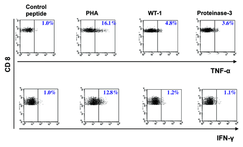
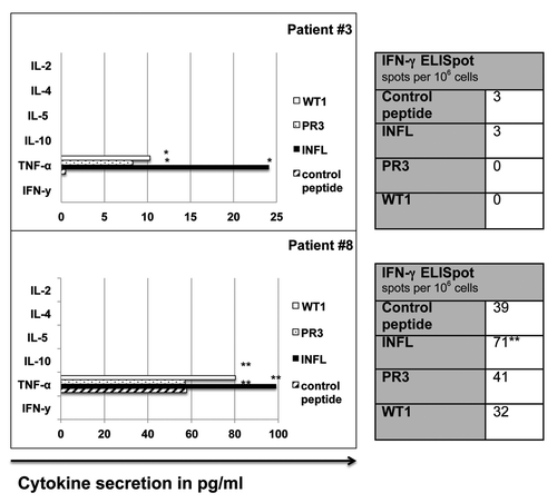
Figure 2. Cytometric bead array (CBA) and IFN-γ ELISpot in patients with TNF-α+/ IFN-γ- CD8+ T cells. TNF-α+/ IFN-γ- antigen-specific T cell responses in patients #3 and #8 as measured by CBA and IFN-γ ELISpot. *, peptide-specific cytokine secretion; **, detectable over background, but not meeting our predefined cut-off for specificity. INFL, Influenza-A; IFN-γ, Interferon-γ; PR3, proteinase-3; TNF-α, Tumor necrosis factor-α; WT1, Wilms´ tumor protein- 1.