Figures & data
Table 1. Cancer immunotherapy based on killing of bacteria-infected tumor cells
Figure 1. RASV increased Ly6-Ghigh MDSCs in the tumor. Two subsets of MDSCs were evident, Ly6-GhighLy6-Cinter cells (upper panel) and Ly6-G interLy6-Chigh cells (lower panel). (A) FACS plot percentages and (B) absolute number of each MDSC subset in the tumor. *p < 0.05. Adapted from Hong et al.Citation15
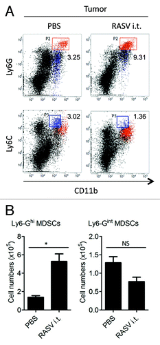
Table 2. Approaches to overcome the immune suppression mediated by MDSCs
Figure 2. Intratumoral injection of RASV induced Ly6-Ghigh granulocytic MDSCs highly secreting TNF-α in the tumor. Tumor-infiltrating cells were stimulated with 200 ng/ml LPS for 2 h, and then, TNF-α secretion by Ly6-GhighLy6-Cinter and Ly6-GinterLy6-Chigh MDSCs was analyzed by intracellular staining. (A) Percentages of TNF-α+ MDSCs in the tumor. (B) The absolute number of TNF-α+ MDSC subsets in the tumor. *p < 0.05. Adapted from Hong et al.Citation15
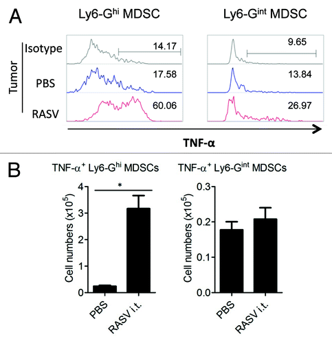
Figure 3. CD4+CD25+FoxP3+ regulatory T-cell levels decreased in tumor-bearing mice after i.t. injection of RASV. (A) The percentages of CD4+CD25+FoxP3+ Tregs among the CD4+ T cell population in splenocytes (n = 6 mice per group). **p < 0.01, ***p < 0.001 compared with naïve control mice. †p < 0.01, Her2/CT26-PBS vs. Her2/CT26-RASV i.t. and Her2/CT26-RASV i.t. vs. Her2/CT26-RASV oral. (B) The percentages of FoxP3+ Tregs among the CD25+ cells are shown after gating the tumor-infiltrating CD4+ T cells. (C) The absolute number of tumor-infiltrating CD4+ T cells and CD4+ CD25+ FoxP3+ Tregs in the tumors. NS, not significant. Adapted from Hong et al.Citation15

Figure 4. Tumor antigen-specific CTL activity and tumor-infiltrating CD8+ T cells. (A) Spleens from RASV-treated mice were obtained, and specific lysis of hP63 (TYLPTNASL) peptide-loaded target cells was estimated by in vivo CTL levels. (B) Results are expressed as the mean cytotoxicity ± SEM from in vivo CTL assays. (C) The absolute number of tumor-infiltrating CD8+ T cells per tumor (left) and per tumor weight (right). *p < 0.05. Adapted from Hong et al.Citation15
