Figures & data
Figure 1. Fluorescence microscopy images of cells. HEK293T cells were transfected with pROP16 (A), pGRA7 (B), pROP16-GRA7 (C), pB7-2 (D), or empty plasmid pEGFP (E). None transfected HEK293T cells (F).
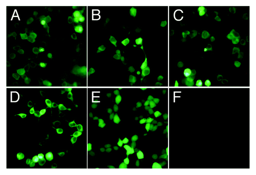
Figure 2. Western blotting analysis of the expression protein in vitro using anti-STAg mouse sera or anti-B7-2 antibody as primary antibody. β-actin serves as a loading control and from the left side there were lysates of HEK293T cells transfected with pEGFP, pROP16, pGRA7, pROP16-GRA7 or pB7-2 recombinant plasmid incubated with the same anti-β-actin antibody.
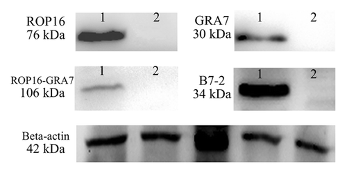
Figure 3. Determination of T. gondii-specific IgGs and IgG subclass titers. (A) Anti-T. gondii IgG titers in sera (diluted 1:100) of mice immunized with pEGFP, pB7-2, pEGFP+pB7-2, pROP16, pGRA7, pROP16-GRA7, pROP16+pB7-2, pGRA7+pB7-2, and pROP16-GRA7+pB7-2. Immune sera were collected at each time point. (B) Anti-T. gondii IgG1 and IgG2a titers in the sera (diluted 1:100) of mice injected with pEGFP, pB7-2, pEGFP+pB7-2, pROP16, pGRA7, pROP16-GRA7, pROP16+pB7-2, pGRA7+ pB7-2, and pROP16-GRA7+pB7-2. Sera were collected at the fourth week post the final injection. Results were expressed as means ± SD for three determinations.
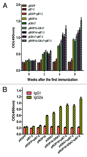
Table 1. Percentages of T lymphocyte subsets from the immunized micea by flow cytometry
Table 2. Cytokine concentrations of culture supernatants of the splenocytes from the immunized micea by ELISA
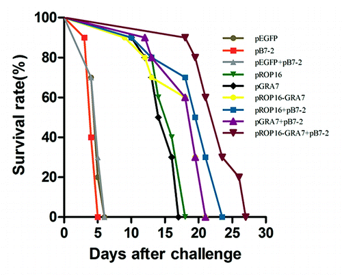
Figure 4. Survival curves of immunized Kunming mice after T. gondii challenge infections. Ten mice per group were challenged with 1000 tachyzoites of the virulent T. gondii RH strain on the 4th week after the final immunization. Mortality was monitored daily for 27 d post-challenge with the parasites.