1,460
Views
25
CrossRef citations to date
0
Altmetric
Case Report

Significant clinical response of progressive recurrent ovarian clear cell carcinoma to glypican-3-derived peptide vaccine therapy

Two case reports

, , , , , , & show all
Pages 338-343 | Received 11 Sep 2013, Accepted 15 Nov 2013, Published online: 21 Nov 2013

Figures & data

Figure 1. (A) Clinical course from the beginning of the GPC3 peptide vaccination. Serum levels of CA125 and CA72–4 decreased after the initiation of therapy. Black arrows indicate vaccinations. The asterisk indicates right inguinal lymph node resection biopsy. The double asterisk indicates bilateral inguinal lymphadenectomy. (B) Contrast-enhanced CT scan showing liver (white, red, blue, and orange arrows) and paraaortic lymph node (yellow arrows) metastases. The size of metastases increased immediately following the initiation of the GPC3 peptide vaccination; however, tumor sizes decreased markedly within three months. (C, D) Pathological findings of primary ovarian carcinoma (C) and right inguinal lymph node biopsy specimens (D). A microscopy image of a hematoxylin and eosin (H&E)-stained section shows CCC (a, i). Immunohistochemical staining for GPC3 and HLA class I showed positivity in the primary ovarian carcinoma, respectively (b, c). Most CCC cells in the resected right inguinal lymph node metastasis appeared to lack GPC3 expression and a reduction in the expression of HLA class I (ii, iii). Immunohistochemical analysis showed a few CD8-positive T cells in the primary ovarian CCC tissue (d), whereas there was little infiltration of CD8-positive T cells in the resected right inguinal lymph node metastasis (iv). Original magnification, x200.
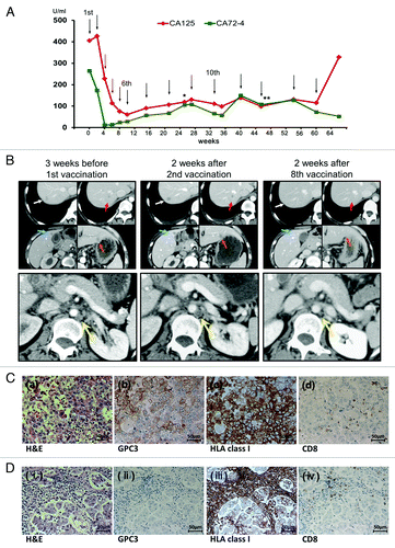
Figure 1. (A) Clinical course from the beginning of the GPC3 peptide vaccination. Serum levels of CA125 and CA72–4 decreased after the initiation of therapy. Black arrows indicate vaccinations. The asterisk indicates right inguinal lymph node resection biopsy. The double asterisk indicates bilateral inguinal lymphadenectomy. (B) Contrast-enhanced CT scan showing liver (white, red, blue, and orange arrows) and paraaortic lymph node (yellow arrows) metastases. The size of metastases increased immediately following the initiation of the GPC3 peptide vaccination; however, tumor sizes decreased markedly within three months. (C, D) Pathological findings of primary ovarian carcinoma (C) and right inguinal lymph node biopsy specimens (D). A microscopy image of a hematoxylin and eosin (H&E)-stained section shows CCC (a, i). Immunohistochemical staining for GPC3 and HLA class I showed positivity in the primary ovarian carcinoma, respectively (b, c). Most CCC cells in the resected right inguinal lymph node metastasis appeared to lack GPC3 expression and a reduction in the expression of HLA class I (ii, iii). Immunohistochemical analysis showed a few CD8-positive T cells in the primary ovarian CCC tissue (d), whereas there was little infiltration of CD8-positive T cells in the resected right inguinal lymph node metastasis (iv). Original magnification, x200.

Figure 2. (A) Clinical course from the beginning of the GPC3 peptide vaccination. Serum levels of CA19–9 and CA125 decreased after the 7th vaccination. The CA19–9 level decreased to within the normal range. Black arrows indicate vaccinations. (B) Plain CT and 18F-FDG PET/CT scans showing retroperitoneal lymph node (white, red, blue and orange arrows) and Virchow's node (yellow arrows) metastases. These metastases were negative on 18F-FDG PET/CT at week 49. (C) Pathological findings of primary ovarian carcinoma. A microscopy image of a hematoxylin and eosin (H&E)-stained section shows CCC (a). Immunohistochemical staining was performed for GPC3, HLA class I, and CD8. (b, c, d). The expression of HLA class I was positive, while that of GPC3 was not, and there was no infiltration of CD8-positive T cells. Original magnification, x200.
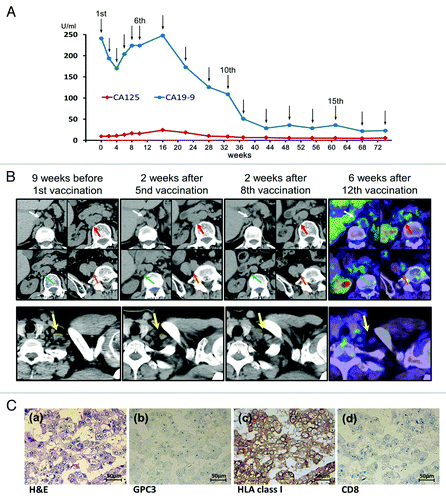
Figure 2. (A) Clinical course from the beginning of the GPC3 peptide vaccination. Serum levels of CA19–9 and CA125 decreased after the 7th vaccination. The CA19–9 level decreased to within the normal range. Black arrows indicate vaccinations. (B) Plain CT and 18F-FDG PET/CT scans showing retroperitoneal lymph node (white, red, blue and orange arrows) and Virchow's node (yellow arrows) metastases. These metastases were negative on 18F-FDG PET/CT at week 49. (C) Pathological findings of primary ovarian carcinoma. A microscopy image of a hematoxylin and eosin (H&E)-stained section shows CCC (a). Immunohistochemical staining was performed for GPC3, HLA class I, and CD8. (b, c, d). The expression of HLA class I was positive, while that of GPC3 was not, and there was no infiltration of CD8-positive T cells. Original magnification, x200.