Figures & data
Figure 1. IL-1β, IL-6, TNF-α, and G-CSF production in PBMCs cultures stimulated with DPT, Hib, PCV7, DPT/Hib, DPT/PCV7, Hib/PCV7, and DPT/Hib/PCV7. PBMCs were obtained from 29 individuals and culture fluids were harvested 24 h after stimulation. Cytokine concentrations were measured using BioPlex 17 cytokine panel. Each bar represents the mean concentration (•) with 95% CI.
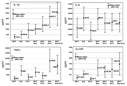
Figure 2. IL-1β production in the PBMCs of 29 individuals. PBMCs were stimulated with DPT, Hib, PCV7, DPT/Hib, DPT/PCV7, Hib/PCV7, and DPT/Hib/PCV7. Columns from left to right in each individual show the production of IL-1β measured by EIA.
Table 1. Number of patients with or without febrile reactions after vaccination with a different combination of vaccines
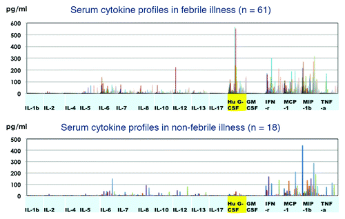
Figure 3. Cytokine profiles of 61 individuals with febrile reactions within 24 h after immunization (upper panel) and those of 18 recipients without febrile illness (lower panel).