771
Views
6
CrossRef citations to date
0
Altmetric
Short Report

Immunotherapy of tuberculosis with Mycobacterium leprae Hsp65 as a DNA vaccine triggers cross-reactive antibodies against mammalian Hsp60 but not pathological autoimmunity

, , , , , , , , , , , , & show all
Pages 1238-1243 | Received 03 Dec 2013, Accepted 15 Feb 2014, Published online: 07 Mar 2014

Figures & data

Figure 1. Production of specific and cross-reacting antibodies against mycobacterial Hsp65, human Hsp60 and DNA after M. tuberculosis infection in mice untreated or treated with DNA-hsp65 immunotherapy. (A) anti-IgG1 and (B) IgG2a antibodies reactive to recombinant M. leprae Hsp65 protein. (C) Detection of antibodies that cross-react with human Hsp60. (D) anti-DNA antibodies. The mean value obtained in healthy non-infected animals in each measurement was subtracted from all the individuals in each group prior to performing statistical analysis. *P < 0.05 compared with all other groups at the same time point by one-way ANOVA and Tukey’s multiple comparisons post-test. F1, female F1 of NZB/NZW mice used as positive controls.
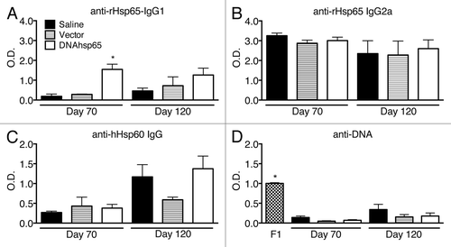
Figure 1. Production of specific and cross-reacting antibodies against mycobacterial Hsp65, human Hsp60 and DNA after M. tuberculosis infection in mice untreated or treated with DNA-hsp65 immunotherapy. (A) anti-IgG1 and (B) IgG2a antibodies reactive to recombinant M. leprae Hsp65 protein. (C) Detection of antibodies that cross-react with human Hsp60. (D) anti-DNA antibodies. The mean value obtained in healthy non-infected animals in each measurement was subtracted from all the individuals in each group prior to performing statistical analysis. *P < 0.05 compared with all other groups at the same time point by one-way ANOVA and Tukey’s multiple comparisons post-test. F1, female F1 of NZB/NZW mice used as positive controls.

Figure 2. Preparation of isotropic and anisotropic organs for paraffin embedding and histological sectioning. Isotropic organs, such as the spleen (A), do not require special procedures and were fixed and embedded in paraffin after their extraction. Anisotropic organs, such as the kidney (B), were pre-sectioned following the orientator method.Citation18 For this method, (1) the organ was cut at random; (2) resultant fragments were cut again with a perpendicular section to the first plane, and (3) step 2 was performed again with the fragments obtained in step 2. After step 3, fragments are considered uniformly isotropic. Then, all of the fragments were immersed in paraffin and sectioned as in A. Grid unit on graph paper equals to 0.5 cm2.
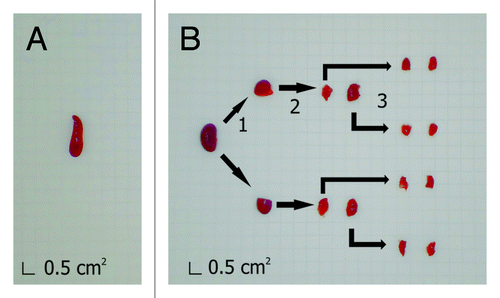
Figure 2. Preparation of isotropic and anisotropic organs for paraffin embedding and histological sectioning. Isotropic organs, such as the spleen (A), do not require special procedures and were fixed and embedded in paraffin after their extraction. Anisotropic organs, such as the kidney (B), were pre-sectioned following the orientator method.Citation18 For this method, (1) the organ was cut at random; (2) resultant fragments were cut again with a perpendicular section to the first plane, and (3) step 2 was performed again with the fragments obtained in step 2. After step 3, fragments are considered uniformly isotropic. Then, all of the fragments were immersed in paraffin and sectioned as in A. Grid unit on graph paper equals to 0.5 cm2.

Table 1. Frequency of minor lesions observed in the histopathology evaluation*

Figure 3. Histopathological analysis of mice infected with M. tuberculosis and treated or not with DNA-hsp65 immunotherapy. Sections of lung, spleen, lymph node, thymus, and liver were obtained from not infected, saline, vector, and DNA-hsp65 groups. The lung samples presented an active inflammatory process with granuloma-like structures across the tissue in samples from saline and vector injected groups. These lesions were less prominent and smaller in DNA-hsp65-treated animals. In contrast, samples from all of the infected groups presented a slight lymphoid hyperplasia in spleen, lymph node, thymus, and liver. The figure shows the results at 120 d after infection. Similar findings were observed at day 70. Tissue was fixed with 10% buffered formalin, and 5 μm sections were stained with hematoxylin and eosin as described in the text. Magnification panels, 100 ×.
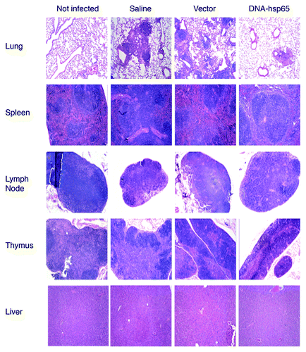
Figure 3. Histopathological analysis of mice infected with M. tuberculosis and treated or not with DNA-hsp65 immunotherapy. Sections of lung, spleen, lymph node, thymus, and liver were obtained from not infected, saline, vector, and DNA-hsp65 groups. The lung samples presented an active inflammatory process with granuloma-like structures across the tissue in samples from saline and vector injected groups. These lesions were less prominent and smaller in DNA-hsp65-treated animals. In contrast, samples from all of the infected groups presented a slight lymphoid hyperplasia in spleen, lymph node, thymus, and liver. The figure shows the results at 120 d after infection. Similar findings were observed at day 70. Tissue was fixed with 10% buffered formalin, and 5 μm sections were stained with hematoxylin and eosin as described in the text. Magnification panels, 100 ×.

Reprints and Corporate Permissions

Please note: Selecting permissions does not provide access to the full text of the article, please see our help page How do I view content?

To request a reprint or corporate permissions for this article, please click on the relevant link below:

Academic Permissions

Please note: Selecting permissions does not provide access to the full text of the article, please see our help page How do I view content?

Obtain permissions instantly via Rightslink by clicking on the button below:

If you are unable to obtain permissions via Rightslink, please complete and submit this Permissions form. For more information, please visit our Permissions help page.