Figures & data
Figure 1. Heat map showed the expression of TLR pathway genes after stimulation with 1 μg/mL CTB at 6 and 12 h. Upregulation was defined as a ≥3-fold increase as compared with negative control, and markedly differentially regulated if the difference was ≥10-fold, and downregulation as a ≤3-fold decrease.
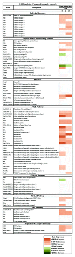
Figure 2. The immune stimulation effect of CTB identified on BMDCs at multiplex levels. (A) Cytokines and chemokines induced 24 h after exposure to CTB. BMDCs were stimulated with either medium or 1μg/mL CTB. Supernatant was harvested 24 h later and analyzed for the presence of cytokines and chemokines according to the manufacturers’ instructions. Expression levels elevated by more than 1.5-fold relative to control were identified as upregulated. Moreover, the time-course and quantitative analysis of cytokines secreted by BMDCs post adjuvants stimulation was evaluated. The concentration levels of IL-6 (B), IL-10 (C), and IL-12 (D) at different time points were evaluated using quantitative ELISA, data from triplicate cultures are shown as mean ± SD *indicates significant difference (P < 0.05) when comparison CTB used with negative or positive control groups.
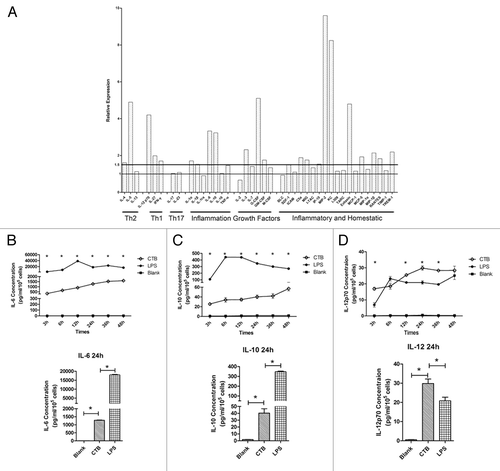
Figure 3. CTB enhanced HIV-1 DNA vaccine cellular immune responses. The immunization regimen is described in . After the last immunization, IFN-γ (A) and IL-2 (B) production of splenic lymphocyte was determined by ELISPOT assay. Notably, CTB strongly enhances the DNA vaccine immunogenicity (P < 0.05). Low dose DNA vaccine co-immunized with adjuvant induced similar Env-specific cell-mediated immune responses level compared with high dose DNA vaccine alone.
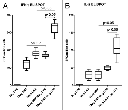
Figure 4. CTB increased HIV-1 Env specific antibody responses at the high DNA dosage. Following the immunization scheme and procedure, the sera were collected to detect HIV-1 Env-specific IgG responses by ELISA assay (A). Anti HIV-1 Env IgG responses induced by 50 μg DNA vaccine co-immunization with CTB were significantly enhanced compared with induction by DNA vaccine alone. (B) IgG isotype analyzed by detecting IgG1 and IgG2a antibody responses in each sample. (C) Avidity of the anti-Env IgG raised by high dose DNA co-administrated with CTB. Sera were analyzed in an HIV Env-specific NaSCN-displacement ELISA. Assays used each serum sample from each group at a dilution of 1:100.
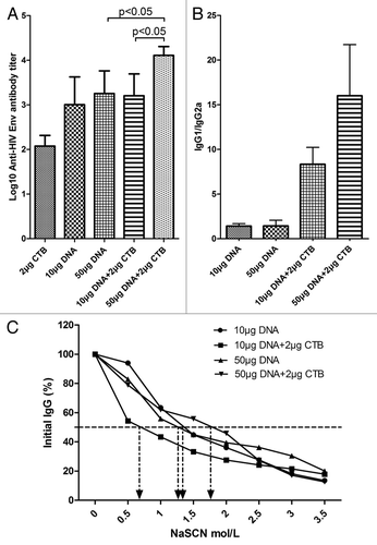
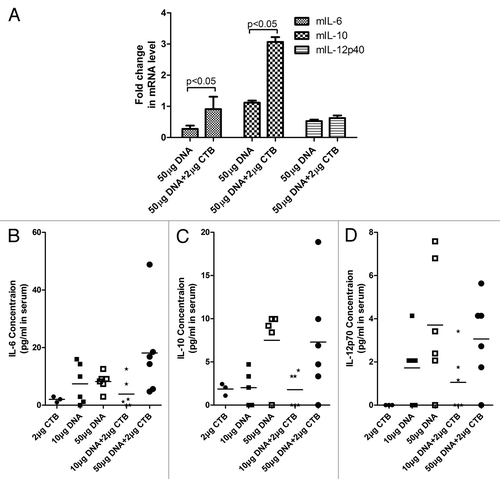
Figure 5. CTB increased the expression level of inflammation cytokines in vivo. (A) IL-6, IL-10, and IL-12 expressions were quantified from administrated mice splenic lymphocyte from group 3 and group 5 mice as detailed in Immunization scheme () and expressed as relative units normalized to GAPDH expression. The reverse transcription reaction was incubated at 48 °C for 30 min, subjected to 10 min initial hot-start activation of the polymerase at 95 °C followed by 40 cycles at 95 °C for 15 s, 60 °C for 1 min. (B), (C), and (D) presents IL-6, IL-10, and IL-12 concentrations in mouse serum by using quantitative ELISA, respectively.