1,380
Views
22
CrossRef citations to date
0
Altmetric
Research Paper

A nonhuman primate toxicology and immunogenicity study evaluating aerosol delivery of AERAS-402/Ad35 vaccine

Evidence for transient t cell responses in peripheral blood and robust sustained responses in the lungs

, , , , , , , , , , , , , & show all
Pages 2199-2210 | Received 17 Jan 2014, Accepted 04 May 2014, Published online: 08 May 2014

Figures & data

Figure 1. Lung histology. Lungs from control (A and B) or vaccinated (C and D) animals were collected on Study Day 18 (A and C) and 43 (B and D). Tissues were sectioned, H&E stained, and examined by microscopy. Arrows with stems indicate perivascular infiltrates. Arrows without stems indicate alveolar macrophages. Bars indicate 100 µm scale.
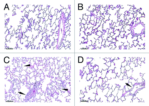
Figure 1. Lung histology. Lungs from control (A and B) or vaccinated (C and D) animals were collected on Study Day 18 (A and C) and 43 (B and D). Tissues were sectioned, H&E stained, and examined by microscopy. Arrows with stems indicate perivascular infiltrates. Arrows without stems indicate alveolar macrophages. Bars indicate 100 µm scale.

Figure 2. BALT histology. Lungs from control (A and B) or vaccinated (C and D) animals were collected on Study Day 18 (A and C) and 43 (B and D). Tissues were sectioned, H&E stained and examined by microscopy. Arrows indicate bronchus-associated lymphoid tissue (BALT). The asterisk indicates the presence of a germinal center. Bars indicate 200 µm scale.
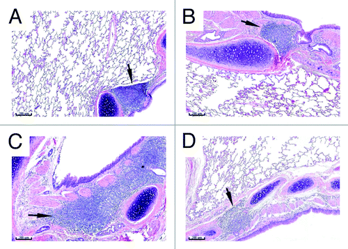
Figure 2. BALT histology. Lungs from control (A and B) or vaccinated (C and D) animals were collected on Study Day 18 (A and C) and 43 (B and D). Tissues were sectioned, H&E stained and examined by microscopy. Arrows indicate bronchus-associated lymphoid tissue (BALT). The asterisk indicates the presence of a germinal center. Bars indicate 200 µm scale.

Table 1. Toxicology results

Table 2. Serum neutralizing antibodies

Figure 3. Serum neutralizing antibodies. Serum was collected from animals prior to study inclusion (pre-bleed) and at each necropsy time point (SD 18 and 42). The Ad35 neutralizing antibody assay was performed and the 90% inhibitory concentration (IC90) calculated for animals receiving three aerosol immunizations on SD 1, 8, and 15 with AERAS-402 (closed circles) or placebo (open circles), or following a single ocular dose of AERAS-402 on study day 1 (triangles). Responses below the limit of detection are reported as an IC90 = 16. Bars represent group median IC90 results.
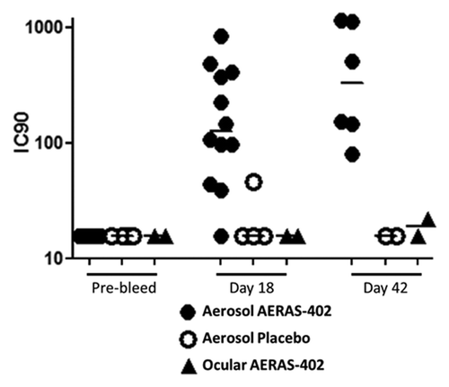
Figure 3. Serum neutralizing antibodies. Serum was collected from animals prior to study inclusion (pre-bleed) and at each necropsy time point (SD 18 and 42). The Ad35 neutralizing antibody assay was performed and the 90% inhibitory concentration (IC90) calculated for animals receiving three aerosol immunizations on SD 1, 8, and 15 with AERAS-402 (closed circles) or placebo (open circles), or following a single ocular dose of AERAS-402 on study day 1 (triangles). Responses below the limit of detection are reported as an IC90 = 16. Bars represent group median IC90 results.

Figure 4. PBMC T cell responses. Animals were immunized with aerosol AERAS-402 or placebo on study days 1, 8, and 15. Blood was collected prior to the first immunization (day 1; A and E) and following the second and third immunizations (days 18 and 43; B, F and C, G, respectively). PBMCs were isolated for the measurement of CD4+ (A-D) and CD8+ (E-H) DMSO-subtracted T cell responses against Ag85A/b and TB10.4 (black and gray bars, respectively) using intracellular cytokine staining for IFN-γ, IL-2 or TNF alone or in any combination. The time course of Ag85A/b-specific T cell responses are shown in plots D and H, for both vaccinated (closed circles) and placebo (open circles) groups. Numbers below bar charts represent unique animal numbers. Circles on dot plots represent individual animal responses. Bars on dot plots represent the median response.
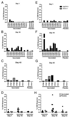
Figure 4. PBMC T cell responses. Animals were immunized with aerosol AERAS-402 or placebo on study days 1, 8, and 15. Blood was collected prior to the first immunization (day 1; A and E) and following the second and third immunizations (days 18 and 43; B, F and C, G, respectively). PBMCs were isolated for the measurement of CD4+ (A-D) and CD8+ (E-H) DMSO-subtracted T cell responses against Ag85A/b and TB10.4 (black and gray bars, respectively) using intracellular cytokine staining for IFN-γ, IL-2 or TNF alone or in any combination. The time course of Ag85A/b-specific T cell responses are shown in plots D and H, for both vaccinated (closed circles) and placebo (open circles) groups. Numbers below bar charts represent unique animal numbers. Circles on dot plots represent individual animal responses. Bars on dot plots represent the median response.

Figure 5. Polyfunctional analysis of PBMC T cell responses on day 18. Animals were immunized with aerosol AERAS-402 (closed circles) or placebo (open circles) on study days 1, 8, and 15. Blood was collected on study day 18 and evaluated by intracellular cytokine staining for production of IFN-γ, IL-2, or TNF for both CD4+ (A) and CD8+ (B) T cells. Circles represent individual responses for each possible cytokine combination. Grey bars represent the group median response.
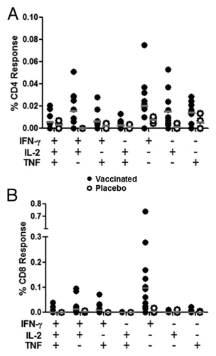
Figure 5. Polyfunctional analysis of PBMC T cell responses on day 18. Animals were immunized with aerosol AERAS-402 (closed circles) or placebo (open circles) on study days 1, 8, and 15. Blood was collected on study day 18 and evaluated by intracellular cytokine staining for production of IFN-γ, IL-2, or TNF for both CD4+ (A) and CD8+ (B) T cells. Circles represent individual responses for each possible cytokine combination. Grey bars represent the group median response.

Figure 6. Lung T cell responses following aerosol immunization. Animals were immunized with aerosol AERAS-402 (black bars) or placebo (white bars) on study days 1, 8, and 15. Two animals each from vaccinated and placebo control animals were pre-selected for BAL collection at the time of necropsy at each necropsy time point (days 18 and 43). Cells were isolated from BAL and CD4+ and CD8+ responses (production of IFN-γ, IL-2, or TNF alone or in combination) evaluated by intracellular cytokine staining following stimulation with Ag85A/b. Bars represent the background-subtracted Ag85A/b response for each animal. Numbers are unique animal identifiers.
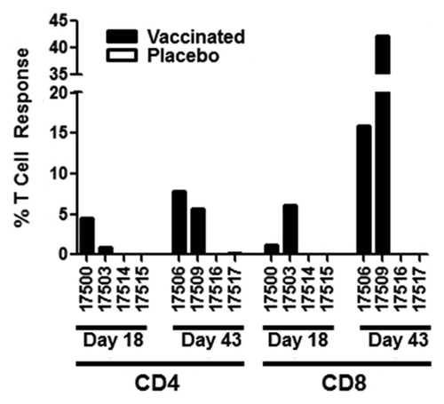
Figure 6. Lung T cell responses following aerosol immunization. Animals were immunized with aerosol AERAS-402 (black bars) or placebo (white bars) on study days 1, 8, and 15. Two animals each from vaccinated and placebo control animals were pre-selected for BAL collection at the time of necropsy at each necropsy time point (days 18 and 43). Cells were isolated from BAL and CD4+ and CD8+ responses (production of IFN-γ, IL-2, or TNF alone or in combination) evaluated by intracellular cytokine staining following stimulation with Ag85A/b. Bars represent the background-subtracted Ag85A/b response for each animal. Numbers are unique animal identifiers.

Figure 7. Polyfunctional analysis of lung T cell responses on study days 18 and 43. Animals were immunized with aerosol AERAS-402 (black bars) or placebo (white bars) on study days 1, 8, and 15. Two animals each from vaccinated and placebo groups were pre-selected for BAL collection at each necropsy time point (days 18 and 43). Cells were isolated and evaluated by intracellular cytokine staining to measure IFN-γ, IL-2, and TNF production by CD4+ (A-B) and CD8+ (C-D) T cells on study days 18 (A,C) and 43 (B,D). Bars represent background-subtracted Ab85A/b responses for individual animals for each of the possible cytokine combinations. Grey bars below plots refer to the functionality shown in the pie charts. Pie charts represent the functionality (3 functions, 2 functions, or one function) of the T cell response for each animal.
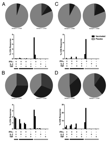
Figure 7. Polyfunctional analysis of lung T cell responses on study days 18 and 43. Animals were immunized with aerosol AERAS-402 (black bars) or placebo (white bars) on study days 1, 8, and 15. Two animals each from vaccinated and placebo groups were pre-selected for BAL collection at each necropsy time point (days 18 and 43). Cells were isolated and evaluated by intracellular cytokine staining to measure IFN-γ, IL-2, and TNF production by CD4+ (A-B) and CD8+ (C-D) T cells on study days 18 (A,C) and 43 (B,D). Bars represent background-subtracted Ab85A/b responses for individual animals for each of the possible cytokine combinations. Grey bars below plots refer to the functionality shown in the pie charts. Pie charts represent the functionality (3 functions, 2 functions, or one function) of the T cell response for each animal.

Table 3. Study design

Supplemental material

Additional material

Download Zip (88.3 KB)

Reprints and Corporate Permissions

Please note: Selecting permissions does not provide access to the full text of the article, please see our help page How do I view content?

To request a reprint or corporate permissions for this article, please click on the relevant link below:

Academic Permissions

Please note: Selecting permissions does not provide access to the full text of the article, please see our help page How do I view content?

Obtain permissions instantly via Rightslink by clicking on the button below:

If you are unable to obtain permissions via Rightslink, please complete and submit this Permissions form. For more information, please visit our Permissions help page.